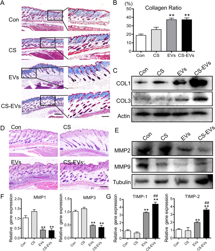
Fig. 6.
Treatment of CS-EVs accelerates skin remodeling. a Histologic images of collagen remodeling by Masson trichrome staining. Scale bar, 150 μm. Boxed areas are shown at higher magnification. b Quantitative statistics of collagen fibers in each group. c Western blot analysis of COL1 and COL3 protein expression. d Histologic images of skin appendage regeneration by HE staining. Scale bar, 100 μm. e Protein expression of MMPs were detected by Western blotting in therapeutic skin tissue. f Gene expression level of MMPs in aging skin. g Gene expression level of TIMPs in aging skin with EVs or CS-EVs treatment was detected by RT-PCR. Data are presented as the mean ± SD. *P < 0.05, **P < 0.01 vs Con; #P < 0.05 vs EVs. All experiments were performed in triplicate

