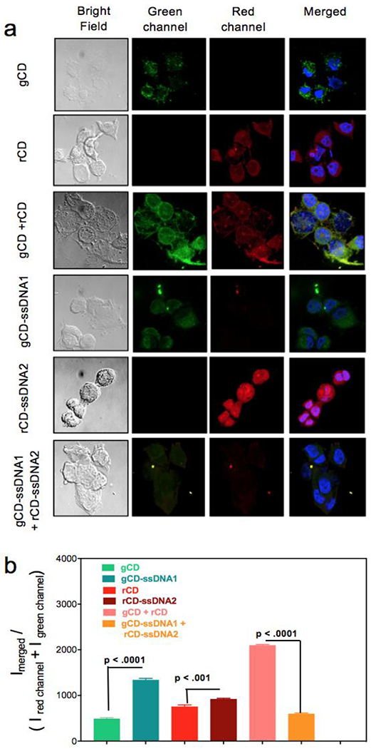
Figure 5.

(a) CLSM images of MCF-7 cells incubated with gCD, rCD, gCD + rCD, gCD-ssDNA1, rCD-ssDNA2 and gCD-ssDNA1 + rCD-ssDNA2. Four different cell panels were acquired, bright field, green channel (for gCD), red channel (for rCD) and overlapped green and blue channels with blue from DAPI. (b) Semi-quantitative fluorescence intensity calculations were performed with help from ImageJ [35] and corroborated with the PL experiments.
