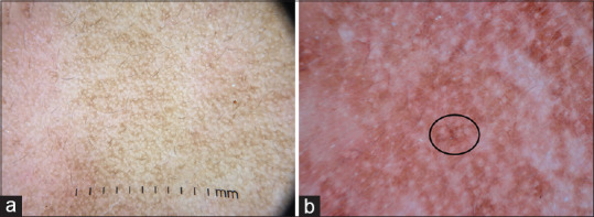
Figure 10.

(a) Dermatoscopy of melasma showing exaggerated pseudoreticular network in comparison to the normal pseudoreticular network in the left half of the image (b) Brownish dots and globules (circle). The appendageal openings are spared. Dermlite DL4 dermatoscope in polarized mode, 10×
