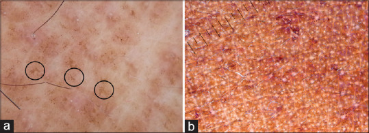
Figure 16.

(a) Dermatoscopy of macular amyloidosis showing a central hub and radially arranged spokes (hub and spoke pattern, circles), Dermlite DL4 dermatoscope in polarized mode, 10× (b) Frictional melanosis showing dispersed pigment dots, Dermlite photo pro dermatoscope in polarized mode, 10×
