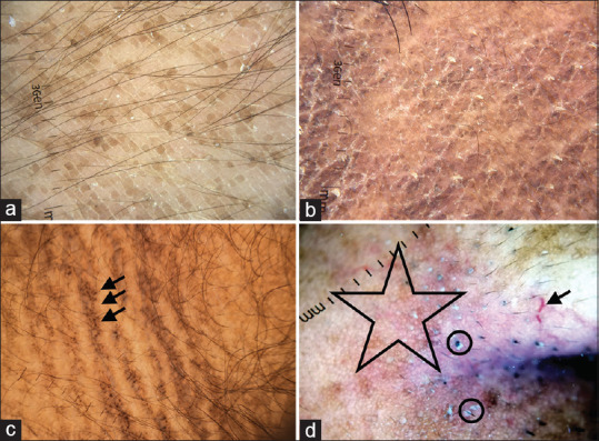
Figure 17.

(a) Terra firma-forme dermatosis showing large brown polygonal scales arranged in cobblestone pattern on dermatoscopy, Dermlite DL4 dermatoscope in polarized mode, 10×. (b) Confluent and reticulate papillomatosis showing brownish homogenous polygonal scales separated by whitish/pale striae arranged in a cobblestone pattern. White fine scales over the surface can be appreciated, Dermlite DL4 dermatoscope in polarized mode, 10×. (c) Acanthosis nigricans shows multiple cristae and sulci. Pigment dots over cristae are characteristic of acanthosis nigricans (arrow), Dermlite photo pro dermatoscope in polarized mode, 10×. (d) Dermoscopy of seborrheic melanosis over nasal ala showing prominent pseudo network (star), ill-focussed vessels (arrow), prominent follicle openings and whitish-yellow excrescences of sebum coating the vellus hair shafts (circle), Dermlite photo pro dermatoscope in polarized mode, 10×
