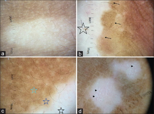
Figure 2.

Dermatoscopy images of vitiligo captured by dermlite DL4 dermatoscope in polarized mode, 10× (a) Diffuse white glow due to complete loss of pigment network at places (b) Stable vitiligo patch showing complete loss of pigment network (star) and well defined hyperpigmented borders (arrows) (c) Progressive vitiligo showing areas of broken pigment network at the edge of the lesion (purple star). This can be contrasted with areas of absent pigment network (black star) and normal reticular pigment network (green star), the three different zones represent the dermatoscopic trichrome sign (d) Reverse pigmentary network (photo courtesy Dr Manoj Nayak, Department of Dermatology AIIMS Rishikesh)
