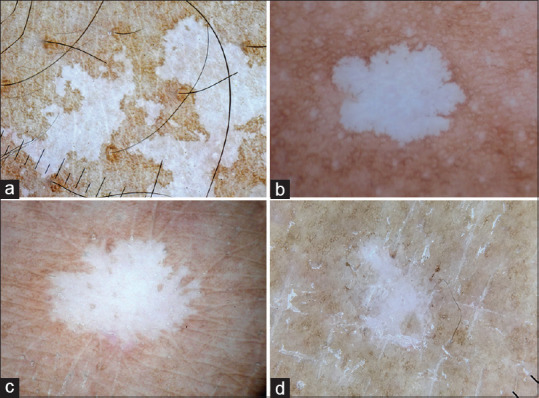
Figure 5.

Dermatoscopy images of idiopathic guttate hypomelanosis showing well-defined margins and complete loss of pigment network (a) Amoeboid pattern with pseudopod like extensions at the margins (b) petaloid pattern with leaf-like extension at the margins (c) Feathery pattern with feather-like striations (d) Nebuloid pattern with indistinct margins. Dermlite photo pro dermatoscope in polarized mode, 10x
