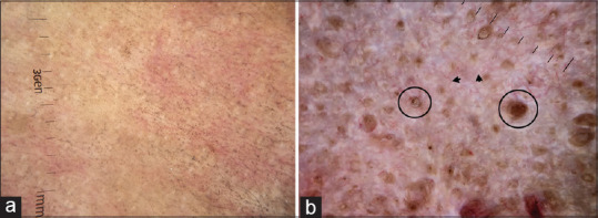
Figure 6.

Dermatoscopic images of lichen sclerosus et atrophicus (a) Early inflammatory phase characterized by background erythema, ill-focused vessels, and peppering of pigment dots (b) Late sclerotic stage characterized by structureless linear white strands representing upper dermal fibrotic bands and follicular plugs (circle). Dermlite DL4 dermatoscope in polarized mode, 10×
