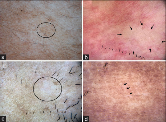
Figure 7.

(a) Dermatoscopic image of nevus depigmentosus showing feathery margins and broken pigmentary network (circle). The diffuse white glow of vitiligo is characteristically missing. (b) Dermatoscopy of nevus anemicus showing focal areas of reduced/absent vasculature (arrows). (c) Dermatoscopic image of progressive macular hypomelanosis showing focal loss of pigment network (circle) and prominent skin markings. (d) Dermatoscopic images of borderline lepromatous leprosy showing focal areas of broken pigmentary network and white chrysalis like structures (arrow). Lesional paucity of appendageal openings and hair follicles can be appreciated. Dermlite DL4 dermatoscope in polarized mode, 10×
