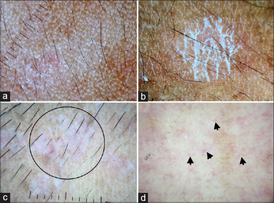
Figure 8.

(a) Pityriasis alba showing focal areas of broken pigment network and diffuse fine scaling, Dermlite photo pro dermatoscope in polarized mode, 10×. (b) Pityriasis versicolor showing accentuated double-edged scales at skin creases, Dermlite photo pro dermatoscope in polarized mode, 10×. (c) Epidermodysplasia verruciformis showing broken pigmentary network, background erythema and ill-focused dotted vessels (circle), Dermlite DL4 dermatoscope in polarized mode, 10× (d) Hypopigmented mycosis fungoides showing areas of broken pigmentary network and spermatozoa like vessels (arrow), Dermlite DL4 dermatoscope in polarized mode, 10×
