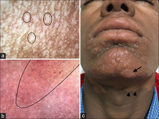
Figure 9.

(a) Dermatoscopic evaluation of patch lesions of post kala-azar dermal leishmaniasis (PKDL) showing loss of pigmentary network and yellowish structureless area representing dermal granulomas (circle). Dermlite DL4 dermatoscope in polarized mode, 10× (b) Dermatoscopy of plaque lesions show yellowish-orange structureless areas and unfocussed vessels representing dermal granuloma, Dermlite DL4 dermatoscope in polarized mode, 10× (c) PKDL involving the muzzle area of the face and neck showing reddish-brown plaques (arrow) and hypopigmented patches (arrowhead)
