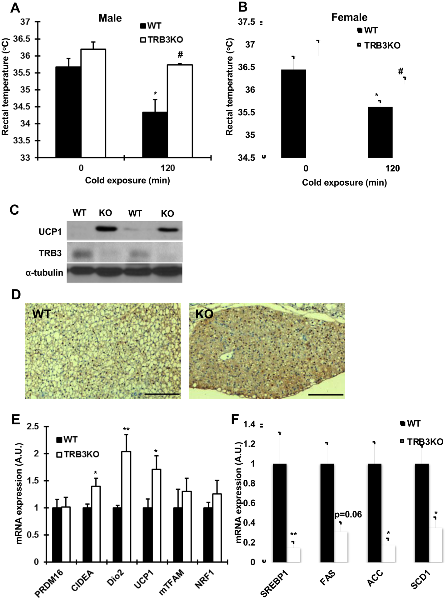
Fig. 2. The functional assessment of brown adipose tissue in TRB3KO mice.

(a,b) Core body temperature was measured in TRB3KO males (a) and females (b) at 4°C (n=5–6). (c) Protein lysates from brown adipose tissue were subjected to Western blot analysis to determine UCP1 expression. (d) Representative histology and UCP1 immunohistochemistry of the brown adipose tissue from wild type (WT) and TRB3KO (KO) mice. Bars indicate 250 μm. We examined 4 independent animals for the analysis. (e,f) Real-time PCR analysis was performed to determine mRNA expression of genes involved in brown adipose tissue differentiation and metabolism (e) and genes involved in lipid metabolism (f) (n=3–8). Data are the means ± S.E.M. * indicates p<0.05 and ** indicates p<0.01 vs. basal or control. # indicates p<0.05 vs. corresponding control.
