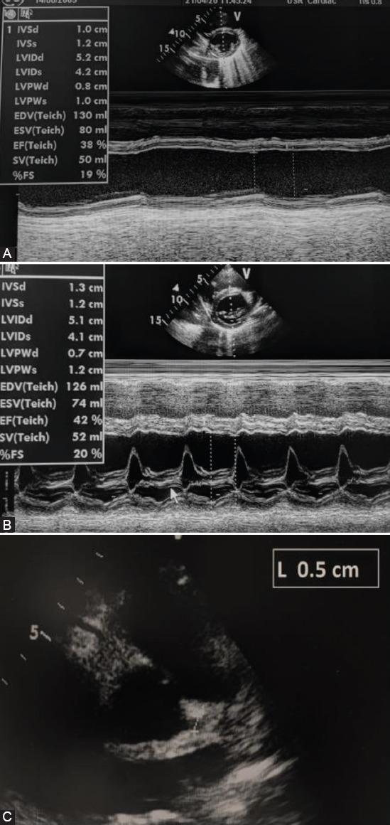
FIGURE 3.

Echocardiography examination at the admission of our patients. (A) 1° patient: Left ventricular function – short axis; normal left ventricular dimensions with an increased wall thickness and decreased ejection fraction of the left ventricle. (B) 2° patient: Left ventricular function – short axis; normal left ventricular and decreased ejection fraction of the left ventricle. (C) 2° patient: Diameter of the left coronary artery (5 mm; Z score +2.77).
