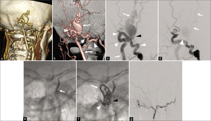
Figure 2:
(a) CT angiography showing the dilated subcutaneous vein suspected the existence of an arteriovenous shunt in the right occipital head. (b) 3D-DSA showing scalp arteriovenous fistula with a fistula at the right occipital head fed by the right OA (arrowhead) and drained to the right OV (arrow). (c) DSA showing the OA and OV has a direct connection at the fistulous point (black arrowhead). (d) TBO using SHORYU HR 4 mm × 7 mm (Kaneka Medix Corp, Osaka, Japan) (arrowhead) placed in the OA just proximal to the fistulous point (arrowhead) reveals that an additional feeder from the STA (arrow) with retrograde flow exists. (e) TAE for STA was performed using coils and NBCA. (f) TAE for dilated OV (arrow) including shunt point (black arrowhead), and OA (arrowhead) using coil and NBCA. (g) The disappearance of the arteriovenous shunt is confirmed. sAVF: Scalp arteriovenous fistula, TBO: temporary balloon occlusion, ST: Superficial temporal artery, OA: Occipital artery, OV: Occipital vein, NBCA: N-butyl-2-cyanoacrylat, DSA: Digital subtraction angiography, CTA: CT angiography.

