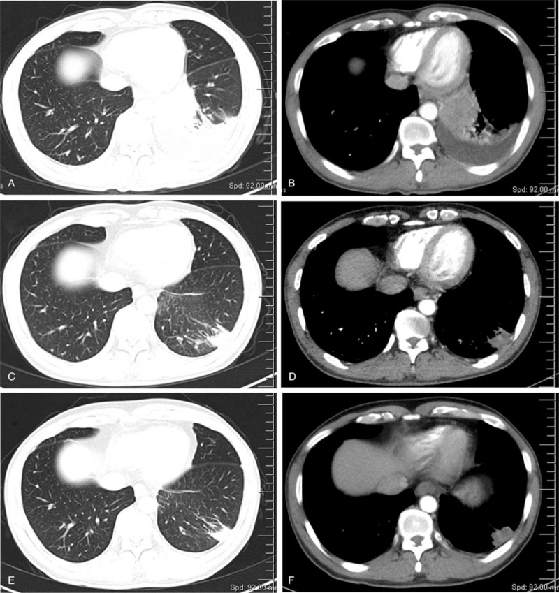
Figure 1.

Chest CT scans of the patient before and after treatment. A-B: First chest CT scan taken on February 13, 2020, note the lesion and pleural effusion in the left lung; C-D: Reexamination of chest CT scan taken on April 20, 2020 after two cycles of treatment; E-F: Reexamination of chest CT taken on June 3, 2020 after four cycles of treatment.
