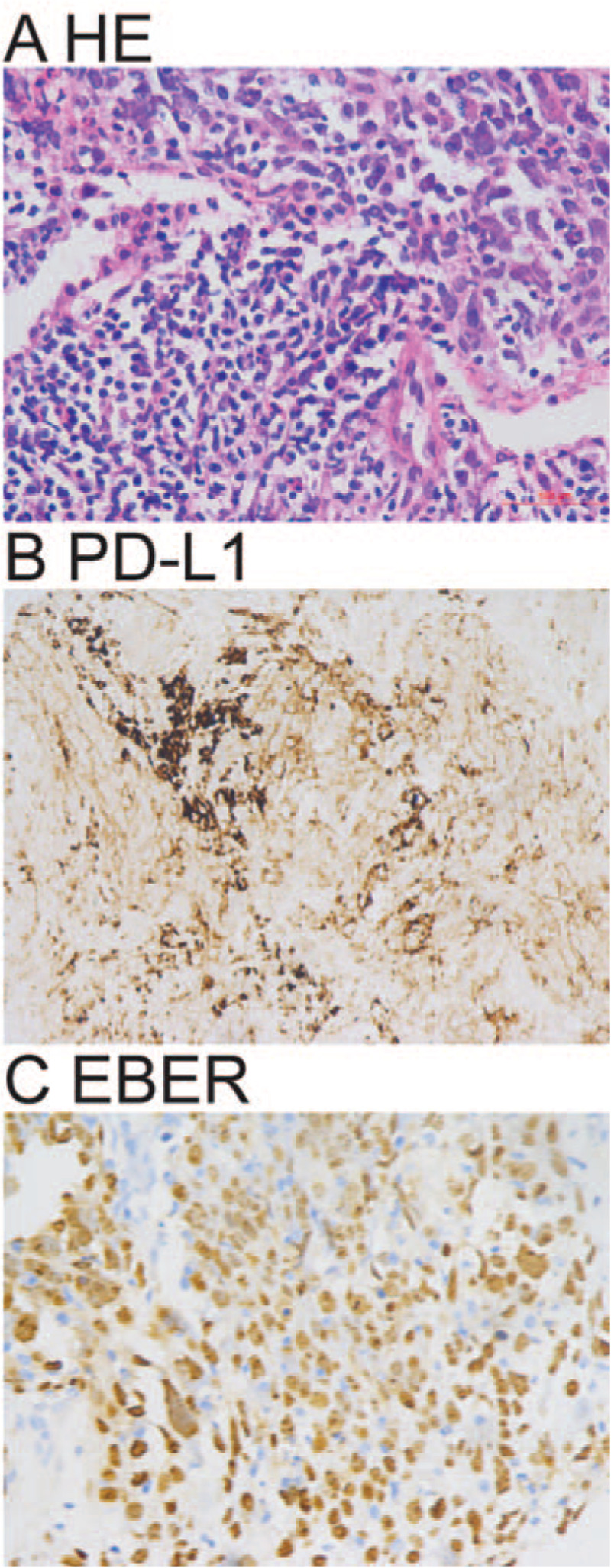
Figure 3.

Percutaneous lung lesion biopsy examination. A. Representative histopathology of percutaneous lung lesion biopsy shows non-small cell carcinoma and lymphoepithelial carcinoma. B. Representative immunohistochemistry image shows PD-L1 (22C3) positive tumor cells. C: Representative in situ hybridization image shows EBER+ tumor cells. HE A × 400; Immunohistochemistry B × 400; in situ hybridization C × 400.
