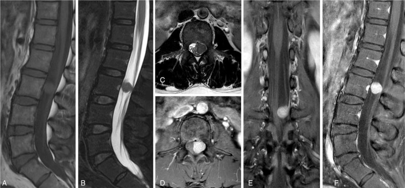
Figure 1.

Preoperative spinal MRI showing a well-circumscribed enhancing mass at the L3 level. The mass, which was isointense on T1- and T2-weighted images (WI), homogeneously enhanced on gadolinium-contrast T1-WI, pushed the cauda equina to the right side, and occupied half the spinal canal volume. (A) Sagittal T1-WI; (B) sagittal T2-WI; (C) axial T2-WI; (D) axial contrast-enhanced image; (E) coronal contrast-enhanced image; (F) sagittal contrast-enhanced image. MRI = magnetic resonance imaging.
