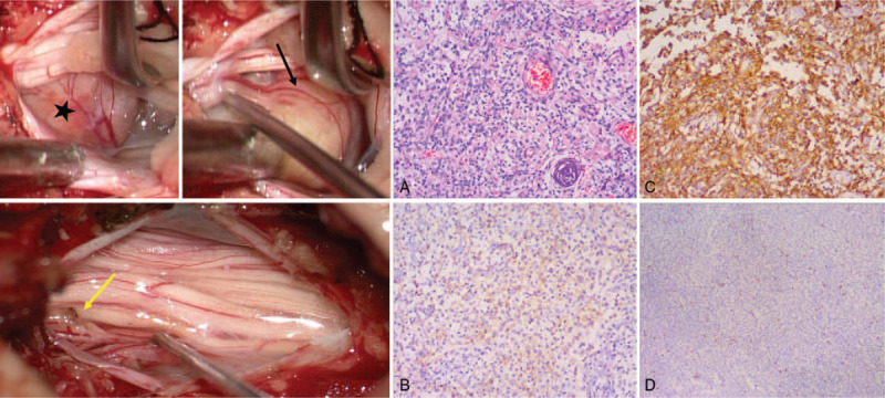
Figure 2.

Intraoperative findings. A yellowish pink, oval, well-encapsulated tumor (marked with a pentagram) adhered to a nerve root (black arrow) without dural attachment. The nerve root was partially removed to achieve complete resection (the nerve stump is indicated with the yellow arrow). Histopathological findings revealed polygonal cells with clear glycogen-rich cytoplasm. (A) Hematoxylin and eosin stain, ×100. (B, C) The immunohistochemical stains showed a positive reaction with EMA (B) and vimentin (C). (D) The Ki-67 index was 20%. EMA = epithelial membrane antigen.
