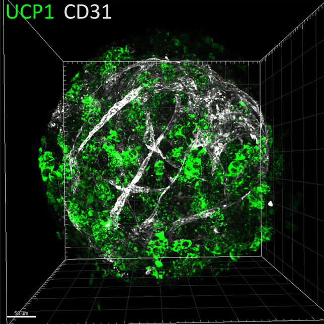
Fig. 2.

Generation of prevascularized hiPSC-3D adipospheres. Adipose progenitors were derived from hiPSCs and induced to differentiate in 3D adipospheres in the presence of endothelial cells. 3D adipospheres were stained for CD31 endothelial cells (white), and UCP1 (green) and then transparized as described by Yao Xi and Dani Christian (Methods in Molecular Biology, iPS cells: Methods and Protocols, in press). Images were acquired on a LSM780 confocal microscope and the 3D reconstruction was generated by the Imaris software. The scale bar is 50 μm
