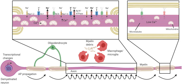
FIGURE 2.
Neuronal adaptations to acute demyelination. Schematic of a partially demyelinated neuron early after demyelination. Conduction is reestablished through the demyelinated segment by the increased expression of sodium channels along the axolemma, but it is notably slower. Demyelinated axons require greater Na+ entry to depolarize the axon, necessitating increased activity of the Na+K+ATPase. Mitochondria increase in number and size within demyelinated axons to meet the higher demand for ATP and also uptake Ca2+. If Na+K+ATPase has sufficient ATP, the NCX is rarely activated in the reverse direction (faded arrows). Transcriptional changes occur within the neuron in response to demyelination and could be critical to these adaptions. Faded text and indicates low activity or levels, bolded text or thick arrows indicates increased levels following demyelination.

