Figure 5.
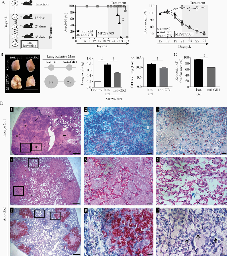
Anti-GR1 treatment reduces tuberculosis severity in MP287/03-infected mice. C57BL/6 mice were infected intratracheally with approximately 100 MP287/03 bacilli and treated with anti-GR1 monoclonal antibodies or isotype control. A, Schematic illustration of the experimental protocol for anti-GR1 therapy, survival curves, and percentages of body weights in relation to time 0. B–D, Mouse lungs were evaluated at day 28 of infection. B, C, Lung macroscopic images, relative masses (circles), lung weights, colony-forming units (CFUs) per lung and morphometric quantifications of the alveolar space are shown. D, Representative lung sections stained using hematoxylin-eosin (D1, D4–D6) or Ziehl-Neelsen (D2, D3, D7–D9) methods. Scale bars correspond to 20 µm (D2, D3, D5, D6, D8, D9) or 500 µm (D1, D4, D7). D1, Extensive areas of pneumonia with numerous foci of caseous necrosis (black stars). D2, Magnified area from D1 (left square) showing alveolitis with numerous intracellular bacilli in the alveoli and extracellular bacilli in region of recent necrosis (white star). D3, Magnified area from D1 (right square) showing low numbers of bacilli in region of central necrosis. D4, Medium-sized lesions surrounded by relatively preserved lung tissue. D5, Magnified lesion from D4 (lower square) showing alveoli filled with homogeneous acellular liquid mass and numerous erythrocytes in alveolar walls. D6, Magnified area from D4 (upper square) showing relatively preserved lung tissue with macrophages phagocytizing erythrocytes (blue arrow). D7, Alveoli filled with extracellular bacilli in medium-sized lesions. D8, Magnified area from D7 (right square) showing numerous extracellular growing bacilli, forming cords and clumps. D9, Magnified area from D7 (left square) showing numerous alveolar macrophages with highly vacuolated cytoplasm (black arrows). Data represent 2 independent experiments with 3–5 mice each. †P < .01; ‡P < .001.

