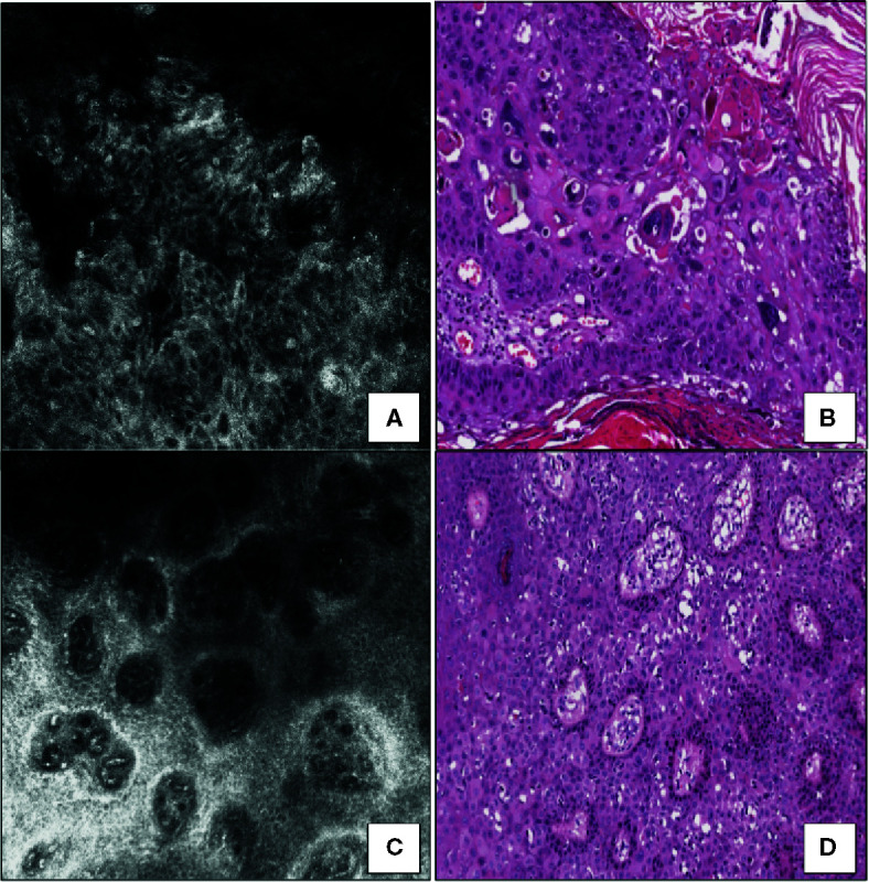
Figure 1.

Squamous cell carcinoma in situ. (A) RCM image at the stratum spinosum showing a marked loss of the normal honeycomb pattern (architectural disarray) due to the presence of markedly variable size, shape, and nuclear morphology keratinocytes. (B) Horizontal histopathology at the same level revealing neoplastic keratinocytes with high-grade nuclear atypia (hematoxylin and eosin; original magnification 400×). (C) RCM image at the dermoepidermal junction showing dilated blood vessels within enlarged edged dermal papillae. (D) Horizontal histopathology at the same level confirming the RCM finding (hematoxylin and eosin; original magnification 100×).
