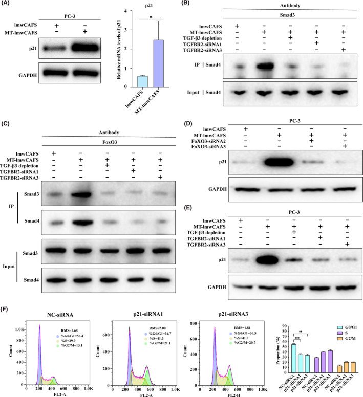
FIGURE 6.

TGF‐β3 from MT‐lmwCAFS activated the FoxO pathway in PC‐3 cells. (A) Western‐blotting and RT‐PCR of p21 in lmwCAFS‐treated PC‐3 cells/MT‐lmwCAFS‐treated PC‐3 cells. (B) and (C) Coimmunoprecipitation analysis of the PC‐3 cells treated with lmwCAFS, MT‐lmwCAFS and MT‐lmwCAFS with TGF‐β3 ligand immune depletion, and the PC‐3 cells with TGFBR2 knockdown and MT‐lmwCAFS treatment. (D) Western blotting of p21 in the PC‐3 cells treated with lmwCAFS and MT‐lmwCAFS, and the PC‐3 cells with FoxO3 knockdown and MT‐lmwCAFS treatment. (E) Western blotting of p21 in the PC‐3 cells treated with lmwCAFS, MT‐lmwCAFS, and MT‐lmwCAFS with TGF‐β3 ligand immune depletion, and the PC‐3 cells with TGFBR2 knockdown and MT‐lmwCAFS treatment. (F) Cell cycle analysis of the PC‐3 cells transfected with p21 siRNA (p21‐siRNA1 and p21‐siRNA3) or negative control siRNA (NC‐siRNA) and treated with MT‐lmwCAFS
