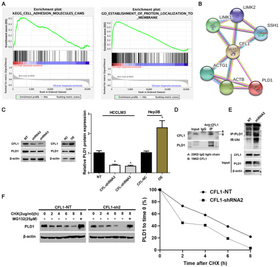
FIGURE 6.

CFL1 regulates PLD1 degradation in HCC cells. (A) KEGG pathway analysis indicated that high expression of CFL1 was closely related to the localization of cell adhesion molecules and proteins located in cell membranes. (B) PLD1 was predicted as a potential CFL1 interactor using the protein interaction database (https://string‐db.org/). (C) HCCLM3 and Hep3B cells were transfected with CFL1 shRNAs (shRNA2 and shRNA3) and CFL1 overexpression plasmid (OE), respectively. Western blotting results indicated that CFL1 positively regulated the PLD1 protein level in HCC cells. (D) Co‐IP assay confirmed the interaction between CFL1 and PLD1 in HCC cells. (E) CFL1 knockdown resulted in increased ubiquitination levels of PLD1 in HCC cells. (F) CFL1 knockdown promoted PLD1 degradation, which could be abolished by MG132 treatment in HCC cells. NT: nontargeting. *p < 0.05
