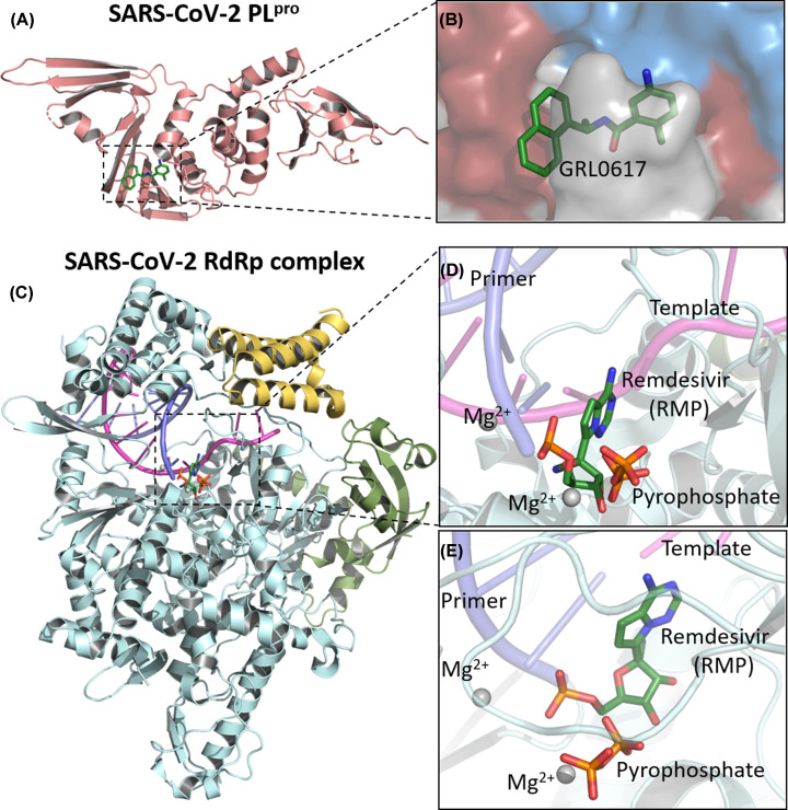
Figure 5. Crystal structure of SARS-CoV-2 PLpro in complex with inhibitor GFL0617 and cryo-electron microscopy structure of the SARS-CoV-2 remdesivir and RNA bound RdRp complex.
(A) Cartoon representation of papain-like protease, PDB ID: 7CMD. (B) An enlarged view of PLpro substrate-binding pocket with GFL0617. (C) Cartoon representation of the nsp12–nsp7–nsp8 RdRp complex with a template-primer RNA and remdesivir. The structure is colored by the elements: nsp12 in pale cyan, nsp7 in pale yellow, nsp8 in bright orange, primer RNA in purple and template RNA in light magenta, PDB ID: 7BV2. (D) Close-up view on the RdRp active site showing the covalently bound remdesivir in its monophosphate form—RMP, pyrophosphate and magnesium ions represented by gray spheres. (E) RdRp binding-pocket in a different view. Images were generated with PyMOL 0.99 [68].

