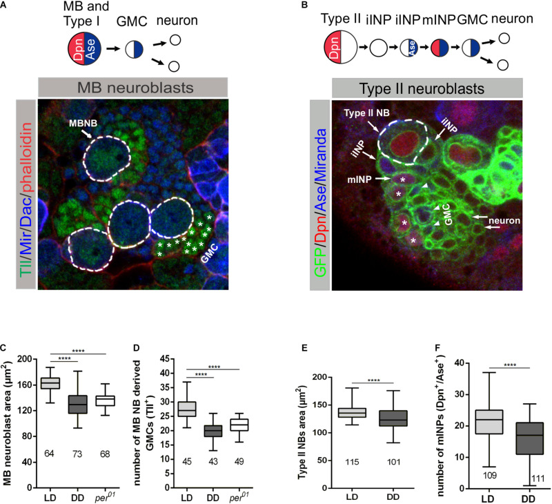
FIGURE 3.
Effect of light and the molecular clock on mushroom body and Type II neuroblast sizes and proliferation. (A) Miranda (Mir blue) was used as a NB marker, Tailless (Tll green) marks mushroom body (MB) NBs and derived GMCs (*), Dachshund (Dac blue) labels MB neurons in wandering 3rd instar larval brains. Phalloidin (red) was used as a marker for cortical actin. (C) The decreased MB NB size under DD conditions and in per01 mutant larvae was accompanied by a reduced number of GMCs (D). (B) Type II NB lineages were marked with GFP (green) expressed under wor-Gal4, ase-Gal80 control. In addition to the cortical NB marker Miranda (blue), nuclear Dpn (red) and Ase (blue) were used to distinguish immature INPs (iINP) and mature INPs (mINP*). (E) Type II NB size as well as the number of mINPs (Dpn+/Ase+) (F) were significantly reduced in larvae under DD conditions. At least 10 brains were analyzed for each genotype or light condition. Data represent the mean obtained from the number of measured NBs or counted progenies ± the max and min size distribution. The number of measured NBs or progeny cells are indicated below of each box plot (****p < 0.00001).

