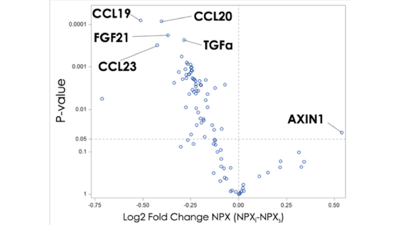
Figure 2.
Inflammatory panel volcano plot illustrating proteomic log2 fold changes in NormalizedProtein eXpression (NPX) in intracranial blood compared with systemic blood. Labeled proteins include C-C motif chemokine 19 (CCL19), C-C motif chemokine 20 (CCL20), fibroblast growth factor 21 (FGF21), transforming growth factor alfa (TGF-α), C-C motif chemokine 23 (CCL23), and axin-1 (AXIN1). Proteins with negative fold change are located to the left of the vertical line and indicate higher expression in systemic blood. Proteins located above the horizontal line are significant (p<0.05). Changes in AXIN1 were found to be non-significant after controlling for the false discovery rate.

