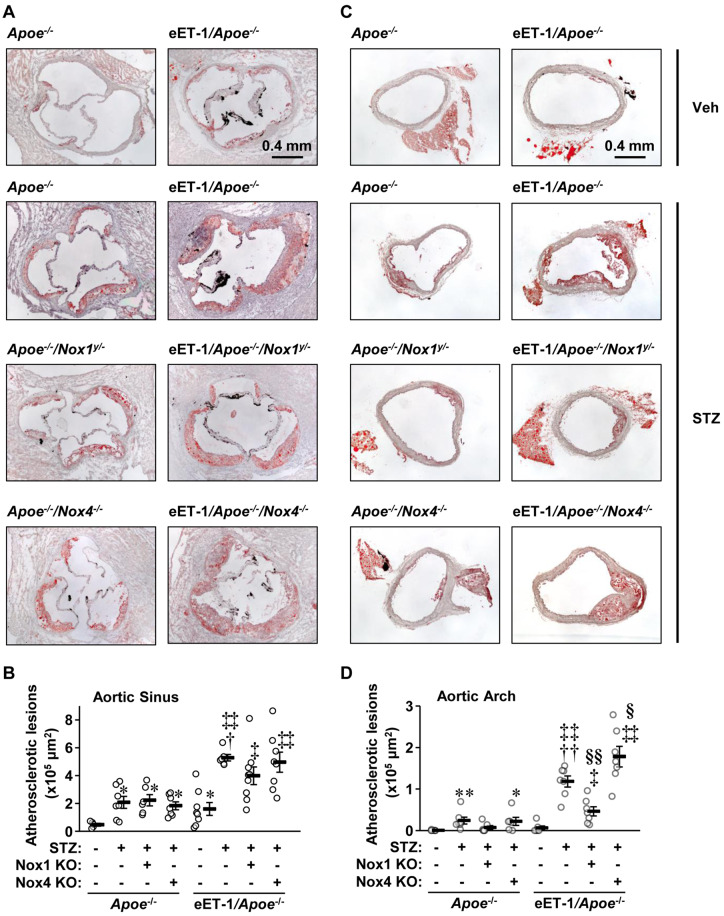
Figure 1.
Endothelial cell-restricted endothelin-1 overexpression worsened atherosclerosis in type 1 diabetes through NOX1. Atherosclerotic lesion areas were determined by oil red O staining in aortic sinus (A, B) and arch (C, D) of Apoe−/−, eET-1/Apoe−/−, eET-1/Apoe−/−/Nox1−/y, and eET-1/Apoe−/−/Nox4−/− mice 14 weeks after intraperitoneal injections with either vehicle (Veh) or streptozotocin (STZ). Representative images of oil red O-stained and Mayer’s haematoxylin-counterstained aortic sinus (A) and arch (C) cryosections are shown. Lesion sizes were calculated as mean μm2 of four sections obtained at 90-μm intervals of aortic sinus or arch. Data are presented as means ± SEM, n = 5–9. Data were analysed using one-way ANOVA followed by a Student–Newman–Keuls post hoc test. *P < 0.05 and **P < 0.001 vs. non-diabetic Apoe−/−, †P < 0.01 and ††P < 0.001 vs. diabetic Apoe−/−, ‡P < 0.01 and ‡‡P < 0.001 vs. non-diabetic eET-1/Apoe−/−, §P < 0.05 and §§P < 0.001 vs. diabetic eET-1/Apoe−/−.

