Figure 3.

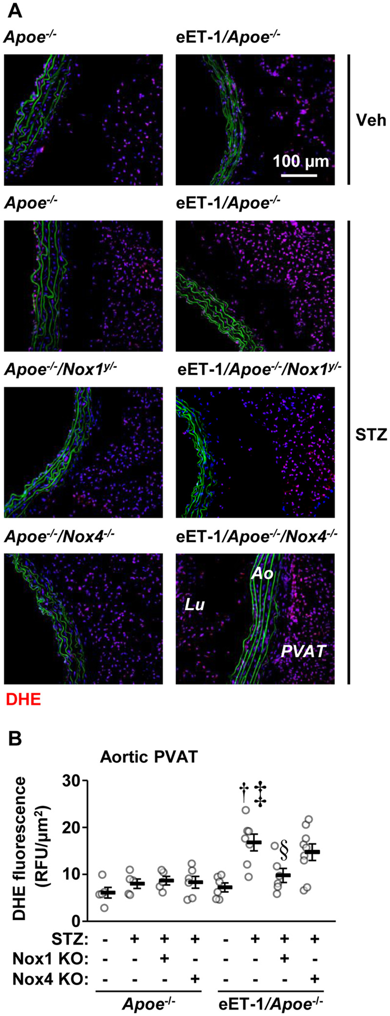
Endothelial cell-restricted endothelin-1 overexpression (eET-1) produced perivascular adipose tissue oxidative stress through NOX1. Reactive oxygen species generation was determined by dihydroethidium (DHE) staining in aortic arch perivascular adipose tissue of Apoe−/−, eET-1/Apoe−/−, eET-1/Apoe−/−/Nox1−/y, and eET-1/Apoe−/−/Nox4−/− mice 14 weeks after intraperitoneal injections with either vehicle (Veh) or streptozotocin (STZ). Representative images of DHE-stained sections are shown in A. Red, green, and blue represent DHE fluorescence, elastin autofluorescence, and 4′,6-diamidino-2-phenylindole fluorescence, respectively. Ao, aorta; Lu, lumen; PVAT, perivascular adipose tissue; RFU, relative fluorescent units. Data are presented as means ± SEM, n = 5–9. Data were analysed using one-way ANOVA followed by a Student–Newman–Keuls post hoc test. †P < 0.001 vs. diabetic Apoe−/−, ‡P < 0.01 vs. non-diabetic eET-1/Apoe−/−, §P < 0.05 vs. diabetic eET-1/Apoe−/−.
