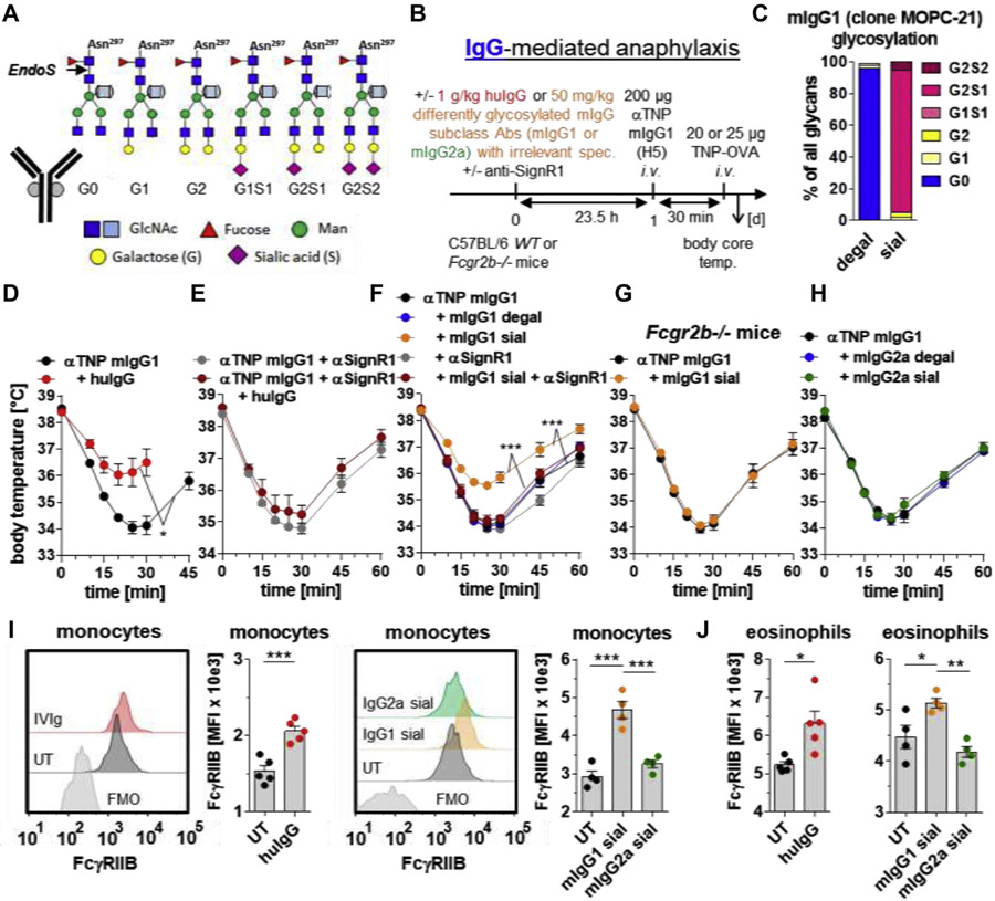
FIG 1.
Enrichment of bulk serum IgG with sialylated murine IgG1, but not IgG2a, with irrelevant specificity attenuates IgG1-mediated anaphylaxis in a SIGN-R1– and FcγRIIB-dependent manner. A, The conserved biantennary N-glycan (4 N-acetylglucosamines [dark blue] and 3 mannoses [green]) at Asn 297 in the IgG Fc part can be modified by fucose (red), bisecting GlcNAc (light blue), galactose (G; yellow), and sialic acid (S; magenta). The cleavage site of EndoS used for IgG glycan analysis is depicted. B, Experimental design of the IgG-mediated 30-minute anaphylaxis model used in the experiments shown in Fig 1, D-H. IgG1-mediated anaphylaxis was induced i.v. with 200 μg of anti (α)-TNP murine (m) IgG1 (clone H5) and subsequent (30 minutes later) i.v. injection of 20 or 25 μg of TNP-OVA. C, Fc glycosylation profiles of in vitro desialylated plus degalactosylated (degal) and galactosylated plus sialylated (sial) mIgG1 antibodies (clone MOPC-21) with irrelevant specificity. D and E, When indicated, intraperitoneal injection of huIgG (IVIg; 1 g/kg) and/or i.v. injection of anti (α)-SIGN-R1 into WT mice. IgG1-mediated anaphylaxis was induced 23.5 hours later. F-H, When indicated, i.v. injection of in vitro galactosylated plus sialylated (sial) or desialylated plus degalactosylated (degal) mIgG1 (clone MOPC-21; 50 mg/kg) or mIgG2a (clone C1.18.4; 50 mg/kg) with irrelevant specificities and/or αSIGN-R1 into (Fig 1, F and H) WT or (Fig 1, G) Fcgr2b−/− mice. IgG1-mediated anaphylaxis was induced 23.5 hours later. The severity of anaphylaxis in all experiments was measured by determining the changes in the body core/rectal temperature on the indicated time points after antigen challenge. n = 4-5 for all groups. I and J, When indicated, intraperitoneal injection of huIgG (IVIg; 1 g/kg) or i.v. injection of in vitro galactosylated plus sialylated (sial) mIgG1 (clone MOPC1; 50 mg/kg) or mIgG2a (clone C1.18.4; 50 mg/kg) with irrelevant specificities into WT mice to analyze FcγRIIB expression (MFI)on blood (Fig 1, I SSC low/CD11b+/F40/80+ classical monocytes and (Fig 1, J) SSC high/GR-1+ (not high) eosinophils by flow cytometry 24 hours later, including overlay histograms of FcγRIIB expression on classical monocytes from representative mice of each group (including FMO controls). Dots represent single mice. Abs, Antibodies; i.v., intravenous/intravenously; MFI, mean fluorescent intensity; OVA, ovalbumin; spec., specificities; temp, temperature; UT, untreated; WT, wild-type.

