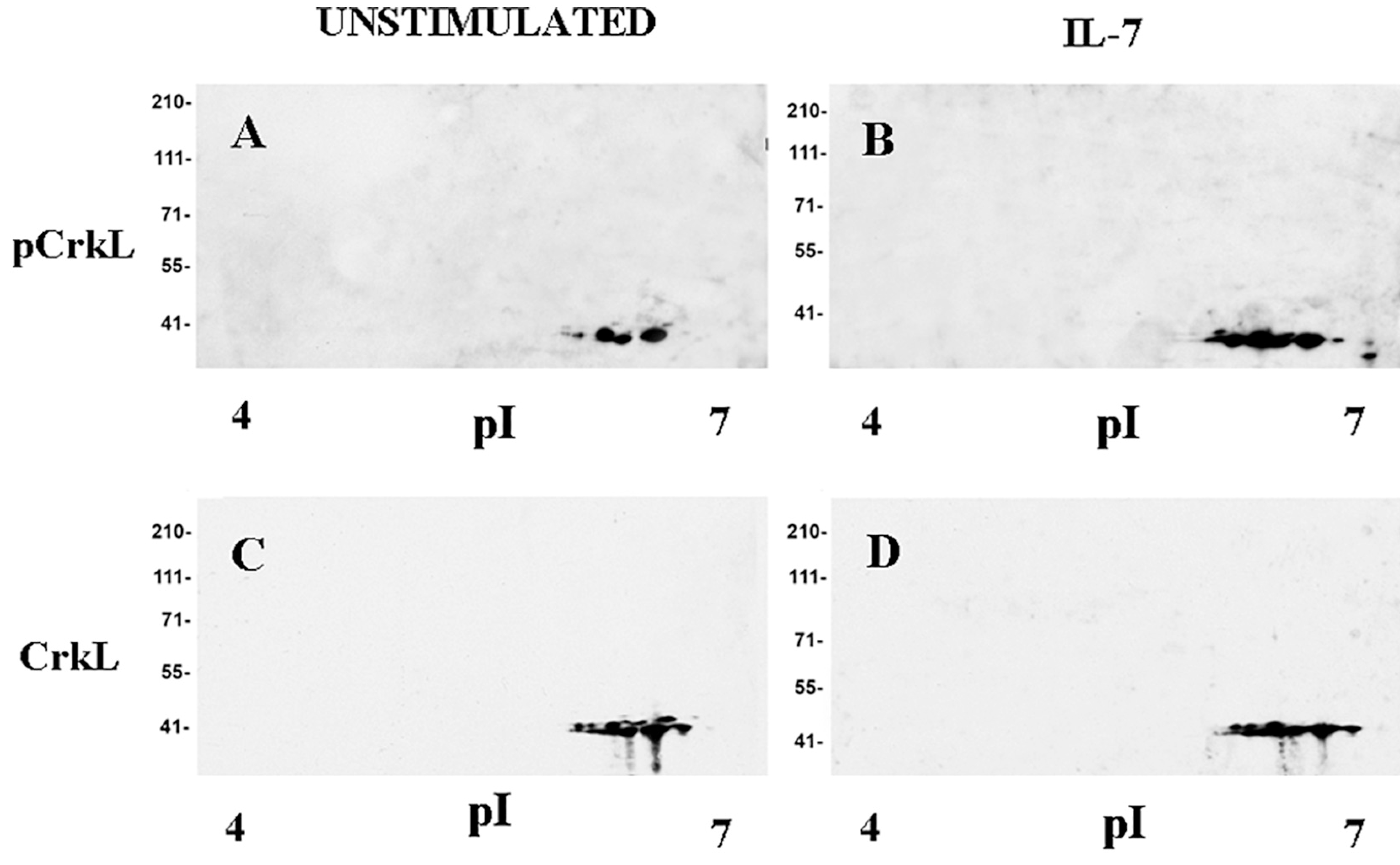
Fig. 2. CrkL tyrosine phosphorylation in IL-7-stimulated D1 cells.

Soluble protein fractions from unstimulated or IL-7-stimulated D1 cells were separated and 2D Western blot analysis was performed as described in Materials and Methods. A, Unstimulated cells, anti-phospho-CrkL antibody; B, IL-7-stimulated cells anti-phospho-CrkL antibody; C, Unstimulated cells, anti-CrkL antibody; D, IL-7-stimulated cells, anti-CrkL antibody. Representative of 3 experiments.
