
Fig. 2.
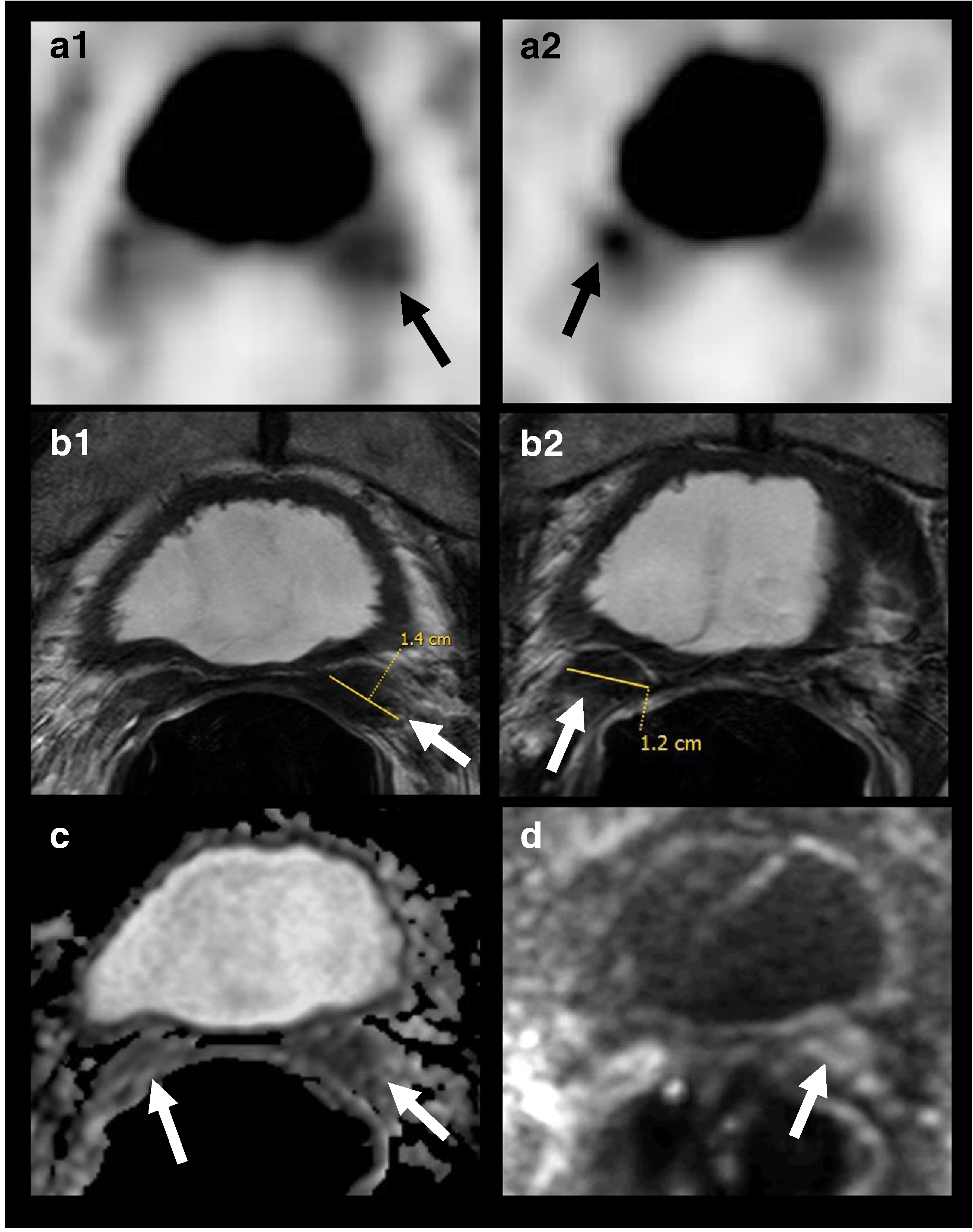
18F–DCFBC PET/CT imaging and mpMRI demonstrating seminal vesicles involvement years after radical prostatectomy: 60-year-old man, with history of prostate cancer, Gleason 7 (4 + 3), status post radical prostatectomy 10 years ago, with pre-scan PSA of 4.7 mg/mL. 18F–DCFBC PET (A1, A2) images demonstrate focal abnormal DCFBC in the bilateral seminal vesicles (arrows), concordant with the MR imaging findings, as seen on T2W MRI (B1, B2), ADC map (C) and b=2000 s/mm2 DW RMI (D); tumor recurrence was confirmed by biopsy, and seminal vesicles surgical resection was performed