
Fig. 4.
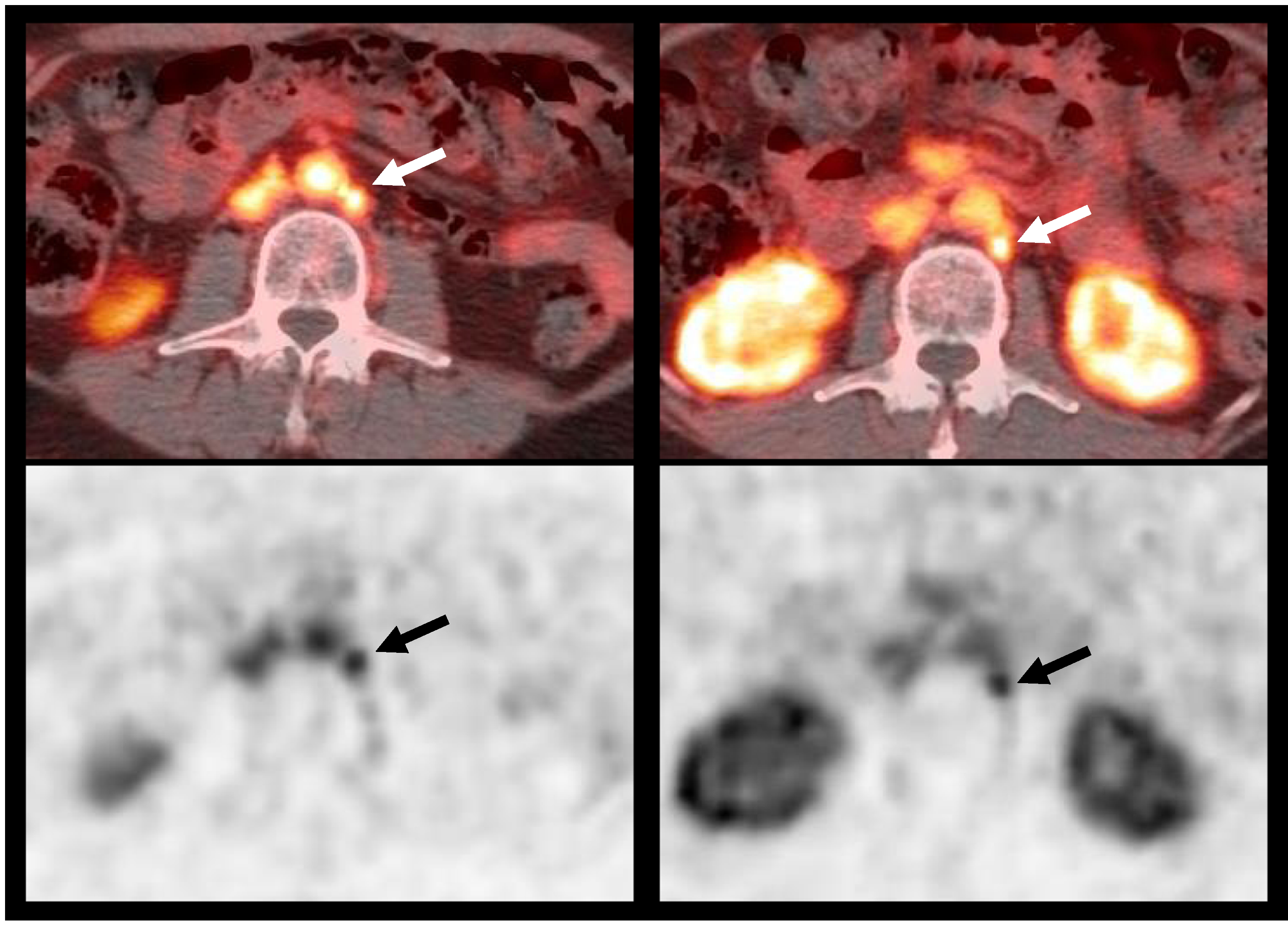
18F-DCFBC PET/CT imaging demonstrating nodal involvement: 61-year-old male, with history of prostate cancer, Gleason 7, status postprostatectomy 2 years ago, with pre-scan PSA of 1.31 mg/mL. 18F–DCFBC fused PET/CT (top) and PET (bottom) images demonstrate two small 9 mm and 7 mm left paraaortic lymph nodes (arrows); biopsy confirmed tumor recurrence. Patient started treatment with enzalutamide.