Figure 1.
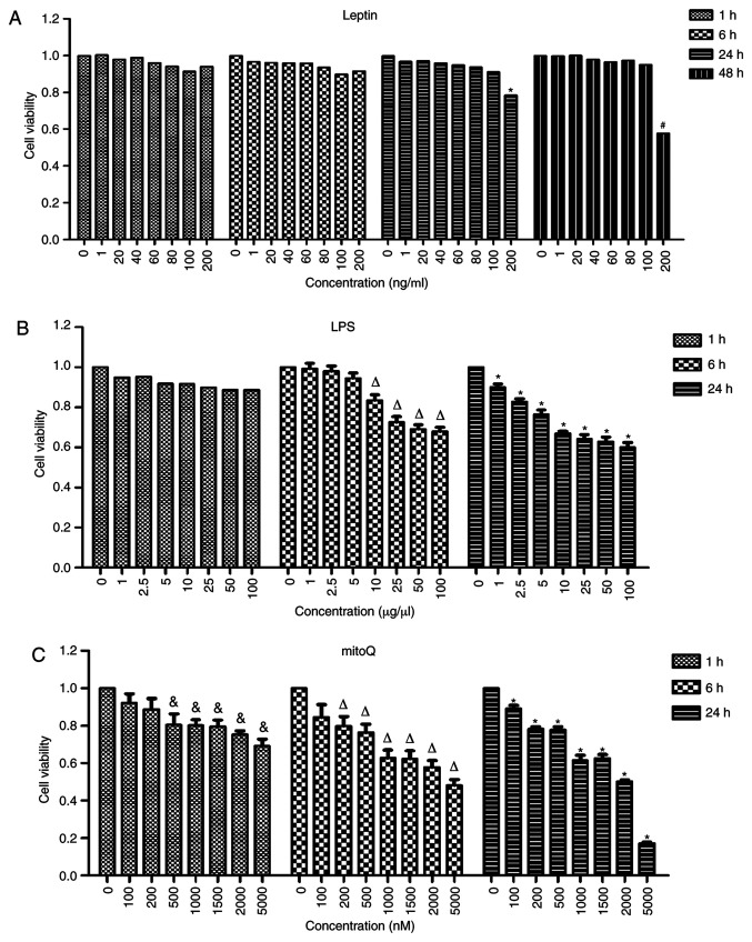
Effects of leptin, LPS and mitoQ on the viability of BEAS-2 cells determined by a CCK-8 assay. (A) Effect of leptin. After 24 h of treatment with leptin at the concentration of 100 ng/ml, the cell viability became significantly lower than that of the untreated cells (P<0.05). (B) Effect of LPS. After 6 h of treatment with LPS at the concentration of 10 µg/µl, the cell viability was significantly decreased compared with that of untreated cells (P<0.05). (C) Effect of mitoQ. The cell viability was significantly decreased after 1 h of treatment with mitoQ at the concentration of 500 nM compared with that of untreated cells (P<0.05). Values are expressed as the mean ± standard error of the mean of 3 samples analyzed in triplicate. #P<0.05 vs. untreated group incubated for 48 h; *P<0.05 vs. untreated group incubated for 24 h; ∆P<0.05 vs. untreated group incubated for 6 h; &P<0.05 vs. untreated group incubated for 1 h. CCK-8, Cell Counting Kit-8; LPS, lipopolysaccharide; mitoQ, mitoquinone.

