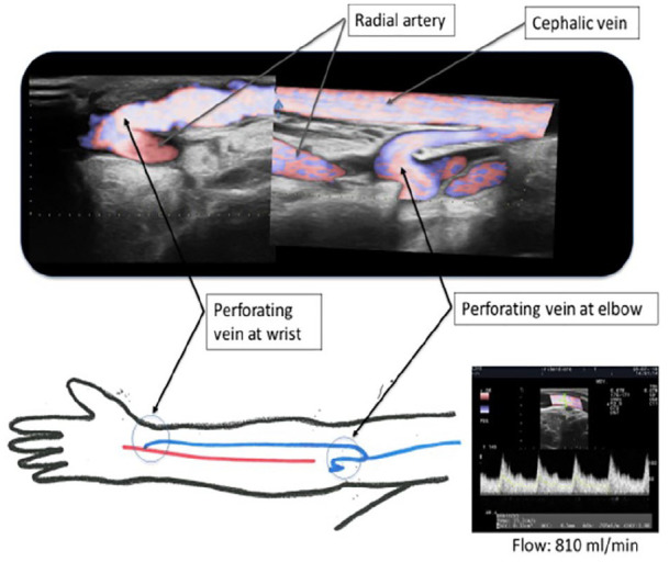
Figure 2.

A composite duplex ultrasound (US) image shows an Ellipsys percutaneous radiocephalic–AVF and a schematic diagram of the radial artery AVF inflow at the wrist with mid-arm outflow through the deep communicating vein (perforating vein) in the cubital fossa. US flow volume of 810 mL/min is documented 6 months following AVF creation.
