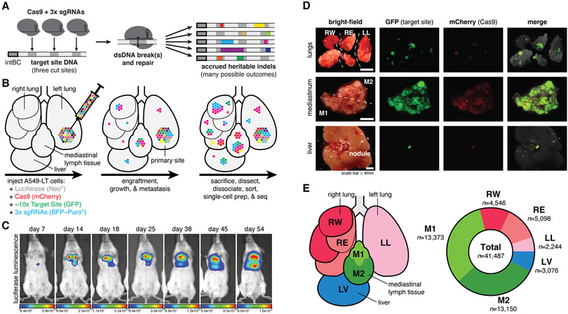
Fig. 1. Lineage tracing in a lung cancer xenograft model in mice.
(A) Our Cas9-enabled lineage tracing technology. Cas9 and three sgRNAs bind and cut cognate sequences on genomically integrated Target Sites, resulting in diverse indel outcomes (multicolored rectangles), which act as heritable markers of lineage. (B) Xenograft model of lung cancer metastasis. Approximately 5,000 A549-LT cells were surgically implanted into the left lung of immunodeficient mice. The cells engrafted at the primary site, proliferated, and metastasized within the five lung lobes, mediastinal lymph, and liver. (C) In vivo bioluminescence imaging of tumor progression over 54 days of lineage recording, from early engraftment to widespread growth and metastasis. (D) Fluorescent imaging of collected tumorous tissues. (E) Anatomical representation of the six tumorous tissue samples (left), and the number of cells collected with paired single-cell transcriptional and lineage datasets (right).

