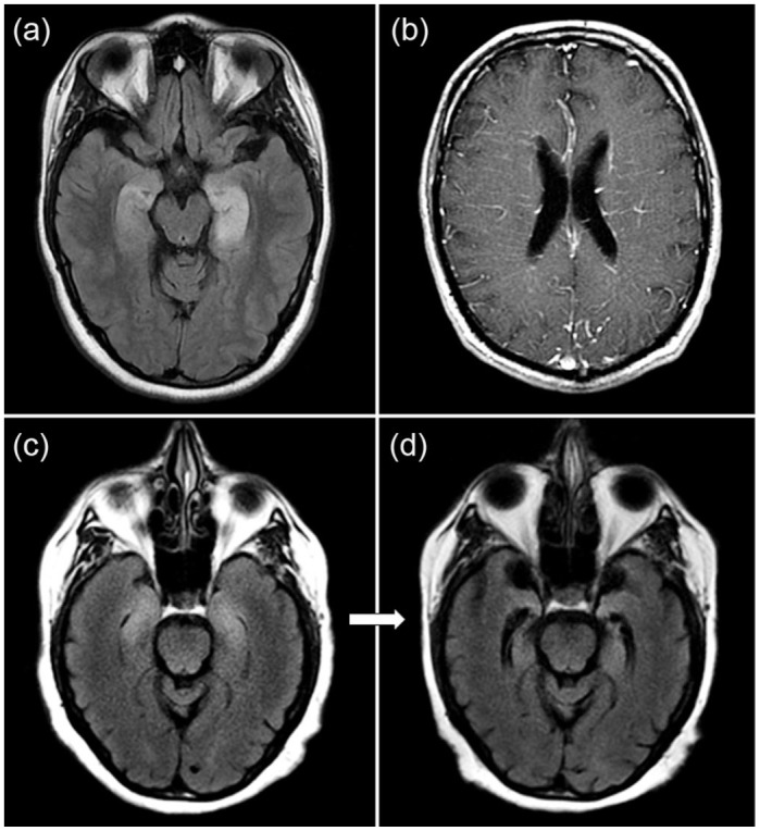
Figure 1.

MRI examples of autoimmune dementia. (a) Bilateral T2-hyperintensities in the mesial temporal lobe are shown in a patient with autoimmune limbic encephalitis associated with GAD65 autoantibodies. (b) Radial perivascular enhancement is shown in a patient with GFAP autoantibodies. (c) Bilateral mesial temporal T2-hyperintensities consistent with limbic encephalitis and occurring after immune checkpoint inhibitor use with an accompanying unclassified neural autoantibody detected with follow-up MRI 1 month later revealing bilateral temporal lobe atrophy (d).
MRI, magnetic resonance imaging.
