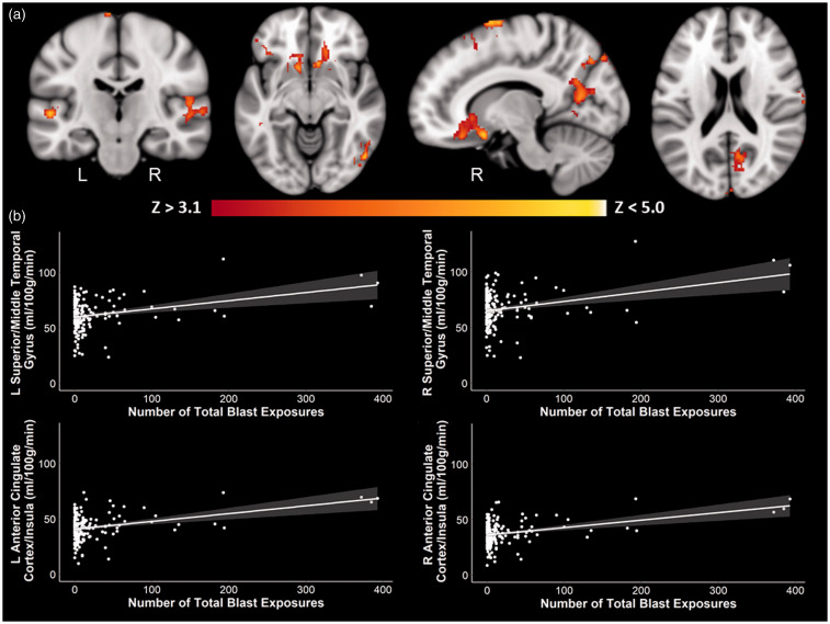
Figure 1.
Positive whole-brain associations between blast exposure and perfusion. There was a significant positive association between the number of total blast exposures and perfusion in regions such as the middle/superior temporal gyri, superior/middle frontal gyri, anterior cingulate cortex, insulae, and occipital cortex. (a) Coronal, axial, and sagittal slices of perfusion in association with the number of total blast exposures are shown in MNI space, controlling for age, scanner, sex, and current PTSD symptom severity. Color scale indicates Z-score threshold. (b) Corresponding scatter plots of selected regions, with 95% confidence intervals shaded in gray, show the positive association between the number of total blast exposures and perfusion. Other brain regions showed similar positive associations as those represented in the scatter plots and are not shown here. L: left; R: right.

