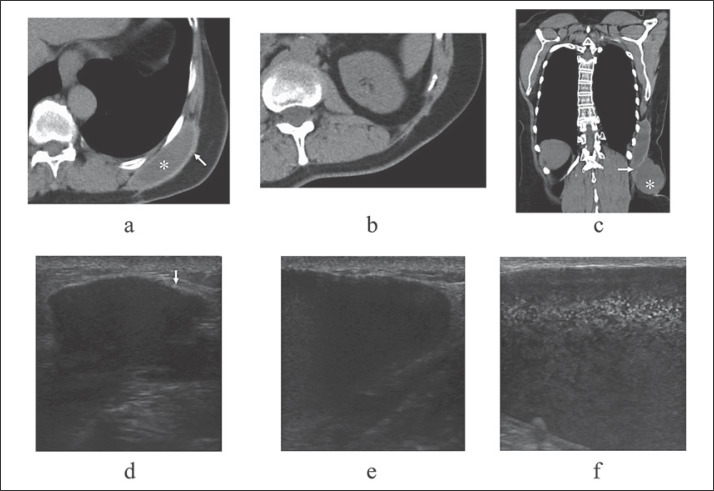
Fig. 2.
Image findings around the seroma. a Computed tomography (CT) showed that the persistent seroma (asterisk) encompassed by thick capsule (arrow) was located just adjacent to the ribs. b CT showed that no apparent protrusion was observed up until 8 years after salvage operation. c Frontal view of the CT showed that the newly protruded presumed seroma (asterisk) was connected to the persistent seroma through the presumed rupture point (arrow). d Persistent seroma with thick capsule (arrow) was observed on ultrasound. e Apparent enlargement of seroma was observed on ultrasound. f Ultrasound showed numerous hyperechoic dots in the seroma.

