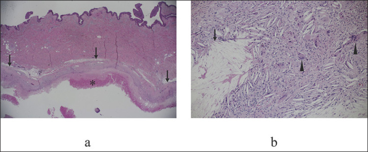
Fig. 3.
Pathological findings. a Low-magnification view showed that the seroma was encompassed by thick hyalinized capsule (arrows) that contained amorphous eosinophilic material (asterisk). b The capsule contained massive cholesterol crystals (arrow) and foreign body multinucleate giant cells (arrowheads).

