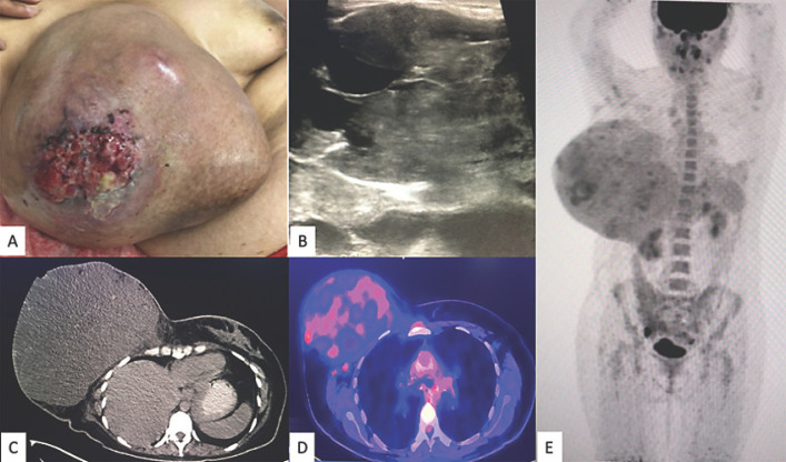
Fig. 1.
A A 40-year-old woman presenting with a giant lesion 40.2 × 36.3 × 15 cm in size in her right breast. B Sonography of the breast showed a huge mass with multiple areas of cystic degeneration alternating with solid tissue, poorly defined margins, irregular vascularity, and thick hyperechogenic septa. C–E18F-FDG PET/CT showed a right breast mass that measured 24.6 × 17.3 cm in the axial plane and 22.5 cm in the cephalocaudal plane, with loss of an interface between it and the pectoralis major muscle, with some hyperdense linear and nodular areas inside, as well as two 9-mm ipsilateral axillary lymphadenopathies.

