Fig. 3.
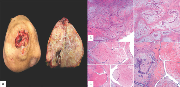
A Surgical specimen. Grossly, the nipple area was extensively ulcerated. The tumor measured 40.2 cm in the greatest diameter. The cut surface presented whitish and yellowish areas. Its heterogeneous consistency was distinguished by prominent firm areas, with other friable and mucoid-like zones. Deeper cystic degeneration was identified. It should be noted that the tumor was entirely lobed, with pushing edges. B Microscopically, the tumor had mixed features. In some areas, the classic pattern of fibroadenoma was evident: ductal epithelial-lined clefts with variable hyperplasia immersed in a paucicellular stroma with a myxoid and collagenized matrix with a lobulated architecture. C These sites alternated with larger leaf-like projections, remarkably hyalinized and with a bland ductal epithelium, consistent with benign phyllodes tumor.

