



Abstract
Chest computed tomography (CT) imaging is a useful tool that provides in vivo information regarding lung structure. Imaging has contributed to a better understanding of COPD, allowing for the detection of early structural changes and the quantification of extra-pulmonary structures. Novel CT imaging techniques have provided insight into the progression of the main COPD subtypes, such as emphysema and small airway disease. This article serves as a review of new information relevant to COPD imaging.
CT abnormalities, such as emphysema and loss of airways, are present even in smokers who do not meet the criteria for COPD and in those with mild-to-moderate disease. Subjects with mild-to-moderate COPD, with the highest loss of airways, also experience the highest decline in lung function. Extra-pulmonary manifestations of COPD, such as right ventricle enlargement and low muscle mass measured on CT, are associated with increased risk for all-cause mortality.
CT longitudinal data has also given insight into the progression of COPD. Mechanically affected areas of lung parenchyma adjacent to emphysematous areas are associated with a greater decline in FEV1. Subjects with the greatest percentage of small airway disease, as measured on matched inspiratory-expiratory CT scan, also present with the greatest decline in lung function.
Introduction
Chronic Obstructive Pulmonary Disease (COPD) is a complex disease with significant heterogeneity in terms of structural and clinical manifestations as well as progression. For instance, subjects with similar severity of airflow obstruction may experience marked differences in symptoms, health-related quality of life, exacerbation prevalence, and exercise capacity. The extent of parenchymal and airway abnormalities, as well as extra-pulmonary manifestations of the disease, also differ between patients with similar levels of spirometric impairment. Chest imaging, particularly computed tomography (CT) and magnetic resonance imaging, provides an in vivo assessment of those processes and enables tangible visualization of COPD pathology. Recent progress in COPD imaging has generated a better understanding of the disease process. This progress has allowed for the improved detection of structural changes in subjects at risk for COPD, the identification of COPD phenotypes, and the additional evaluation of associated extra-pulmonary structures. For example, imaging has demonstrated that airways are not only remodeled but are also completely lost.1,2 Airway loss has been shown to precede the development of emphysema, and subjects with the highest airway loss on CT, also experience the greatest decline in lung function.3,4
A myriad of factors have contributed to this progress in COPD imaging, including the increased use of CT scanning; improved computational capabilities; artificial intelligence-based development of algorithms for imaging; and the increased availability of large cohorts of subjects with imaging characterization, such as the COPDGene Study,5 Evaluation of COPD Longitudinally to Identify Predictive Surrogate End-points (ECLIPSE),6 Subpopulations and Intermediate Outcome Measures of COPD Study (SPIROMICS),7 and Canadian Cohort Obstructive Lung Disease (CanCOLD) Study.8
In this review, we have primarily focused on CT imaging as a modality since it is most widely used, and it provides a tremendous source of information. Three main aspects of imaging are explored: a) imaging of subjects at risk for and with mild COPD, b) imaging of extra-pulmonary structures, and c) imaging of COPD progression. Recent reviews on other imaging modalities and aspects of COPD are available.9,10
Imaging Of Subjects At Risk For And With Mild COPD
The diagnosis of COPD is primarily based on spirometric measures of airflow obstruction (i.e., a ratio of forced volume in one second to forced vital capacity [FEV1/FVC] <0.7);11 however, substantial clinical manifestations, such as dyspnea, chronic bronchitis, exacerbations, and hospitalizations, as well as structural changes, can occur before susceptible individuals meet the criteria for airflow obstruction.12,13,14,15
Lung parenchyma imaging
Imaging studies suggest that smokers undergo a radiographic bi-phasic remodeling process, with a more predominant diffuse parenchymal inflammation in mild disease followed by emphysematous destruction in more advanced stages. In a large cohort, investigators demonstrated that CT lung mass (i.e., lung parenchyma Hounsfield Units [HU] +1,000) was higher in smokers with mild disease (i.e., Global Initiative for Obstructive Lung Disease [GOLD] stage 1) than those without COPD and those with GOLD stages 2–4.16 Although the findings of that study may reflect the effect of cigarette smoking on increasing lung density rather than inflammation, additional studies in smokers have linked high-density lung areas with clinical outcomes and inflammation biomarkers. Investigators explored areas of normal-looking parenchyma and examined their local density to elucidate their potential clinical implications.17 They then determined the percentage of lung occupied by normal-looking parenchyma with density values at the percentile 95th or above (abbreviated: NormHA)— taken from the distribution of local histograms of density in never-smoking subjects. The investigators found that smokers with the highest quartile of NormHA had lower exercise capacity and a higher risk of death than did those in the lowest quartile, independent of emphysema and interstitial lung abnormalities.17 They also found that a higher percentage of regions of lung occupied by NormHA was related to higher levels of C-reactive protein, a biomarker of systemic inflammation. Although the study still lacks histological validation, this CT metric has the potential to identify mild lung tissue injury (or inflamed tissue), before the phase of lung destruction (emphysema) becomes apparent.
CT is a non-invasive, in vivo method to identify and quantify emphysema. Emphysema is quantified as percent of low attenuation areas (%LAA) below a selected HU threshold, typically −950 on inspiratory CT scans, and gas trapping is defined as %LAA below −850HU on expiratory CT scans. Emphysema is typically thought of as a manifestation of overt COPD, however, in a large CT study, 26% of smokers who do not meet the spirometric criteria for COPD had radiographic evidence of lung disease, encompassing emphysema (defined as >5% of LAA on inspiratory CT) or gas trapping (defined as >20% of LAA on expiratory CT), suggesting that spirometry underestimates the effects of cigarette smoking in this population.12 Moreover, in the general population, the emphysema-like lung was demonstrated to be predictive of incident airflow limitation.18,14 This information could prove useful for clinicians, as increased emphysema on CT scans in smokers at risk to develop COPD may warrant closer monitoring and earlier follow-up.
Airways imaging
CT imaging has also demonstrated that in never-smoker subjects, the smallest central airways (i.e., airways visible on clinical CT scans) are associated with the lowest lung function, and central airways in smokers with COPD are smaller than in smokers without COPD, suggesting an interplay of native tracheobronchial structure and environmental exposures such as tobacco smoke.19,20,21 One possible consequence of this interplay is that people with smaller central airways may be more prone to develop airflow obstruction when exposed to tobacco smoke. Moreover, people who have small central airways relative to lung size —a phenomenon called dysanapsis— are more likely to develop COPD over time.22 In terms of bronchial structural changes, symptomatic current and former smokers without COPD had higher central airway-wall thickening compared to their asymptomatic counterparts.14
Central airways may reflect small airway (i.e., <2mm lumen diameter) pathology, the site of airflow obstruction in COPD. A surrogate measure of small airway disease is air trapping on expiratory CT images. Computational advances have made it possible to match voxel-to-voxel inspiratory and expiratory images, a technique called Parametric Response Mapping (PRM)23 (Figure 1). Based on lung density thresholds described above, PRM classifies the paired voxels as normal, emphysema (PRMEMPH), and functional small airway disease (PRMfSAD), the latter representing non-emphysematous air trapping areas of the lung. The metric has been recently validated against microCT measures of small airway disease in lung tissue from COPD patients who underwent a lung transplant.24 Specifically, a higher percentage of PRMfSAD was related to lower circularity and luminal area as well as complete obstruction of terminal bronchioles; a higher PRMEMPH was correlated with larger air space size, lower alveolar surface area, and fewer alveolar attachments per terminal bronchioles. In COPD pathogenesis, small airway disease is thought to precede the emphysematous destruction of the lung. Interestingly, a study in smokers without meeting criteria for COPD, showed that higher PRMfSAD was associated with a higher residual volume to total lung capacity ratio (RV/TLC), an index of air trapping; a lower transfer factor for carbon monoxide adjusted for alveolar volume; and spirometric measures of small airways.25 These findings suggest that high PRMfSAD might be an early indicator of pulmonary pathology in this population.
Figure 1:
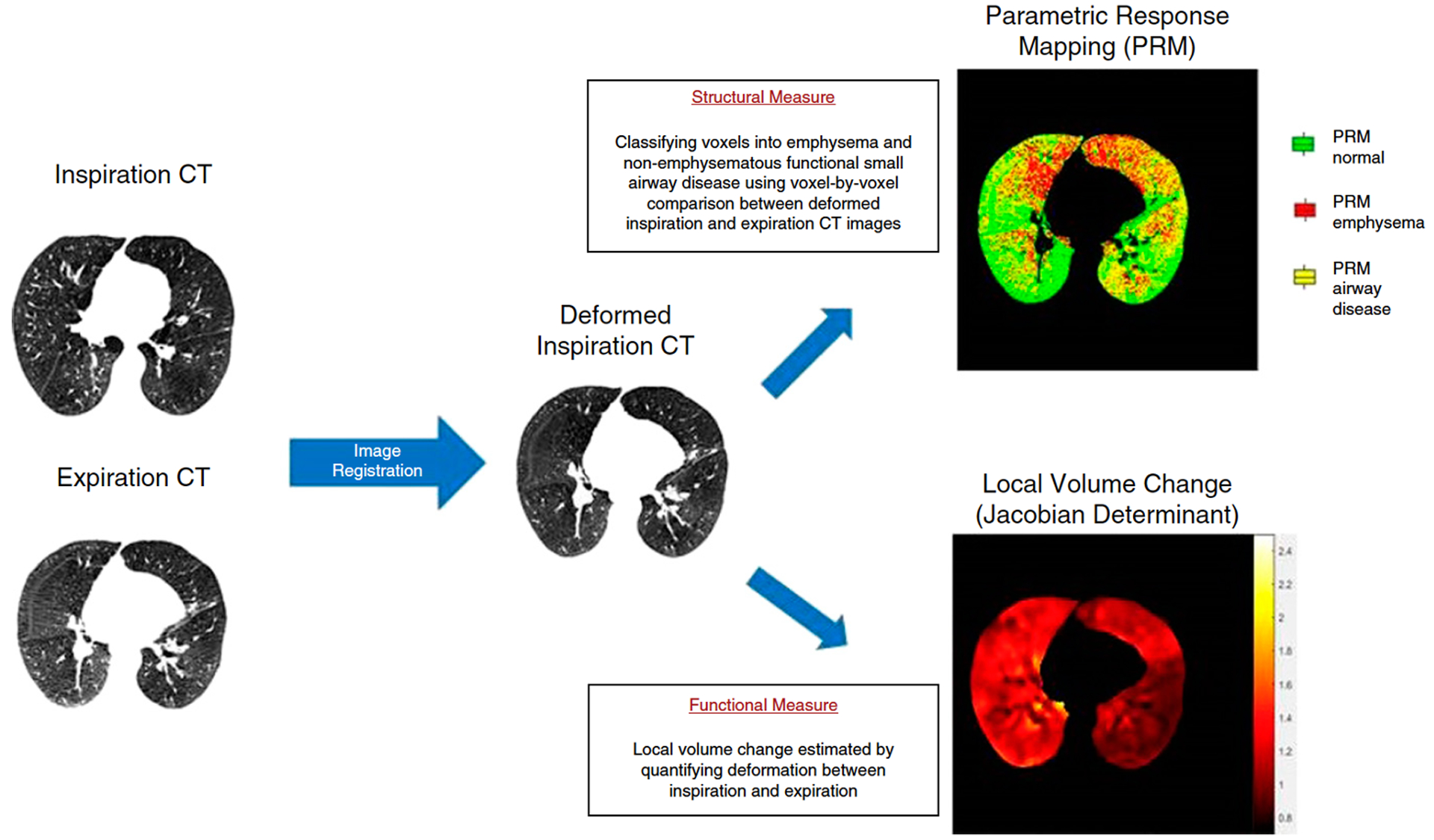
The technique of image registration and two fundamental approaches to extracting clinically relevant information from image registration. Inspiratory and expiratory images are matched voxel-by-voxel. Structural measures can be obtained by performing a voxel-by-voxel anatomic comparison and assessing the corresponding computed tomography (CT) density change from expiration to inspiration, with compensation for lung deformation through image registration. The top right shows a representative axial section with localization of emphysema (red), functional small airways disease (yellow), and normal (green) voxels in a patient with moderate chronic obstructive pulmonary disease (COPD). The bottom right depicts functional changes on the same slice. Here, the amount of lung deformation between inspiration and expiration is used to derive a measure of regional ventilation, termed the Jacobian determinant, a measure of local volume change from full inspiration to end expiration. The Jacobian determinant ranges from 0 to infinity; values greater than 1 indicate local expansion, and values less than 1 indicate local contraction. PRM = parametric response mapping.
Reprinted with permission of the American Thoracic Society. Copyright © 2020 American Thoracic Society. Annals of the American Thoracic Society 2018; 15(3): 281–289
CT imaging has also helped demonstrate variations in central airway tree branching, which has been identified in over one-quarter of the general population.26 Investigators have shown that central airway branch variants have strong familial aggregation and increased COPD susceptibility, particularly among smokers. They also found that one airway branch variant – the absence of the right medial-basal airway – was associated with single nucleotide polymorphisms of fibroblast growth factor 10 —FGF10, which is involved in a broad range of cellular processes including, lung morphogenesis— supporting a genetically programmed airway structure-function endotype.26 COPD pathology is characterized by a profound loss of small airways as shown on microCT, up to 40% in mild-to-moderate COPD, and up to 80% in severe-to-very severe COPD when compared to smoker controls. The loss of normal parallel pathways in this setting may explain the increased (4 to 40 fold) resistance to airflow, a central pathophysiologic feature of the disease.1,27 The loss of small airway branches seems to extend to central airways as well, as demonstrated in patients with advanced and mild-to-moderate disease.2,3 In a Canadian study, the total airway count (i.e., the number of branches counted on a CT-based bronchial tree) of subjects with mild-to-moderate COPD and never-smoking controls and smokers without COPD (Figure 2) was compared. The authors found that patients with COPD had a 19% and 17% lower total airway count compared to never-smoking subjects and smokers without COPD, respectively.3 Furthermore, parent airways with missing daughter branches had smaller lumens and thinner walls than those parent airways without missing branches, which raises the possibility that airways shrink before disappearing. The loss of airways also raises the question as to whether it may have therapeutic implications, particularly for inhalation therapy.28
Figure 2:
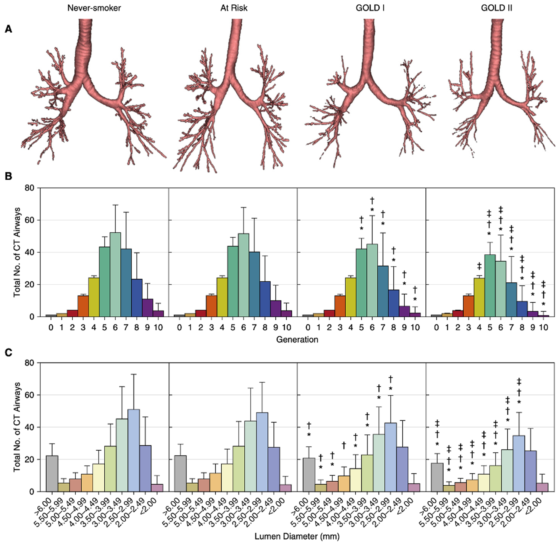
Computed tomography airway count by generation and airway lumen diameter. The three-dimensional reconstruction of the segmented airway tree generated by VIDA Diagnostics Inc. for never-smokers and participants at risk and with Global Initiative for Chronic Obstructive Lung Disease (GOLD) I and GOLD II chronic obstructive pulmonary disease (A). The plot summary data show airway counts for airways color coded by airway generation (B) and by various sizes divided into discrete bins (C). Error bars represent the SD of the airway counts for all participants. *Significantly different from neversmoker. †Significantly different from at-risk. ‡Significantly different from GOLD I. CT = computed tomography.
Reprinted with permission of the American Thoracic Society. Copyright © 2020 American Thoracic Society. American Journal of Respiratory and Critical Care Medicine 2018; 197(1): 56–65.
Imaging of Extra-pulmonary Manifestations of COPD
COPD is considered a systemic disease affecting multiple organs. Advances in imaging have allowed for the identification and quantification of extra-pulmonary structures, leading to a better understanding of their relationships with the lungs and the potential clinical implications. Large vessels such as the pulmonary artery and aorta, as well as the heart, skeletal muscles, and bones, can be segmented and quantified; and their associations with clinical outcomes determined.
Large vessels
Pulmonary hypertension is frequent in patients with COPD and linked to worse outcomes.29 Although the standard method to diagnose pulmonary hypertension is right heart catheterization, imaging modalities such as echocardiogram and CT are non-invasive alternatives to identify this condition.29 Enlargement of the main pulmonary artery (PA) has long been recognized as an imaging feature of pulmonary hypertension. On CT images, PA enlargement is measured as the ratio of the diameter of PA to the aorta (PA:A) on a single axial slice. It has been demonstrated that a PA:A >1 is a better indicator of pulmonary hypertension than echocardiogram in subjects with severe COPD.30 This advantage of CT scanning might be due to the poor acoustic windows for the echocardiogram resulting from hyperinflation in severe COPD. Subjects with a higher PA:A or PA:A ratio >1 (vs. those with ratio <=1) had greater right ventricular (RV) end-diastolic and end-systolic volume indices as well as lower RV ejection fraction, as measured with cine cardiac MRI and echocardiogram.31,32 PA:A ratio was also related to PA systolic pressures and pulmonary vascular resistance. Imaging data also has given greater insight into the pulmonary circulation in smokers. Non-contrast CT studies have shown that the enlargement of the main pulmonary artery is associated with the loss of distal pulmonary vessels, measured as the volume of vessels <5 mm2 —a measure called BV5. The PA:A ratio correlated inversely with total intra-parenchymal pulmonary blood vessel volume and BV5.31 This CT measure also provides prognostic information. In a study using two large cohorts of COPD subjects, a PA:A ratio >1 was independently associated with COPD exacerbations, suggesting that vascular factors might be considered as a potential etiology of exacerbation episodes.33 Since cardiovascular disease accounts for approximately fifty-percent of deaths among COPD patients, investigators have also evaluated the relationship between pulmonary artery enlargement and mortality. Several studies have consistently shown that the PA:A ratio is related to increased mortality.34–36 These findings suggest that PA:A ratio provides clinically relevant information, is a useful marker of pulmonary vascular changes and has prognostic value.
Heart
Cardiac dysfunction is frequent in COPD and is thought to be due to tobacco smoke exposure, hyperinflation, and pulmonary vascular remodeling. Both enlargements of the RV (i.e., cor pulmonale), as well as decreased RV filling and reduced-end diastolic chamber (i.e., cor pulmonale parvus), are manifestations of cardiac dysfunction described in COPD.37,38 To understand the complex relationships between the heart, the pulmonary vasculature, and emphysema, as well as their associations with clinical outcomes, investigators used numerous imaging techniques (Figure 3). Using mathematical models and cardiac models as a reference, they segmented and quantified the heart in non-contrast CT scans.39 This CT method provides epicardial (i,e., myocardium volume plus chamber volume) measures of the RV and left ventricle volumes. The generated CT measurements were correlated with echocardiogram and MRI measurements, imaging modalities typically used in clinical settings to evaluate the heart.40 The technique was applied to a large cohort of smokers. The investigators found that RV was smaller in subjects with GOLD stage 4 vs. stage 1 COPD and that higher arterial pruning (i.e., lower BV5) and emphysema on CT were each independently associated with greater RV sizes.39 In further analysis, however, arterial vascular pruning was related to greater RV size only in subjects with mild airflow obstruction and in those with a low burden of emphysema (i.e., <9.1%). RV enlargement was inversely associated with exercise capacity only among subjects in the lowest quartile of emphysema. A greater RV size was also related to increased mortality. However, the effect was modified by arterial BV5, such that death risk was 63% higher in patients with RV enlargement and arterial pruning, with no significant increased risk for death without pruning.39 Altogether the CT findings on heart, pulmonary vasculature, and lung density suggest a vascular phenotype in COPD that warrants further investigation.
Figure 3:
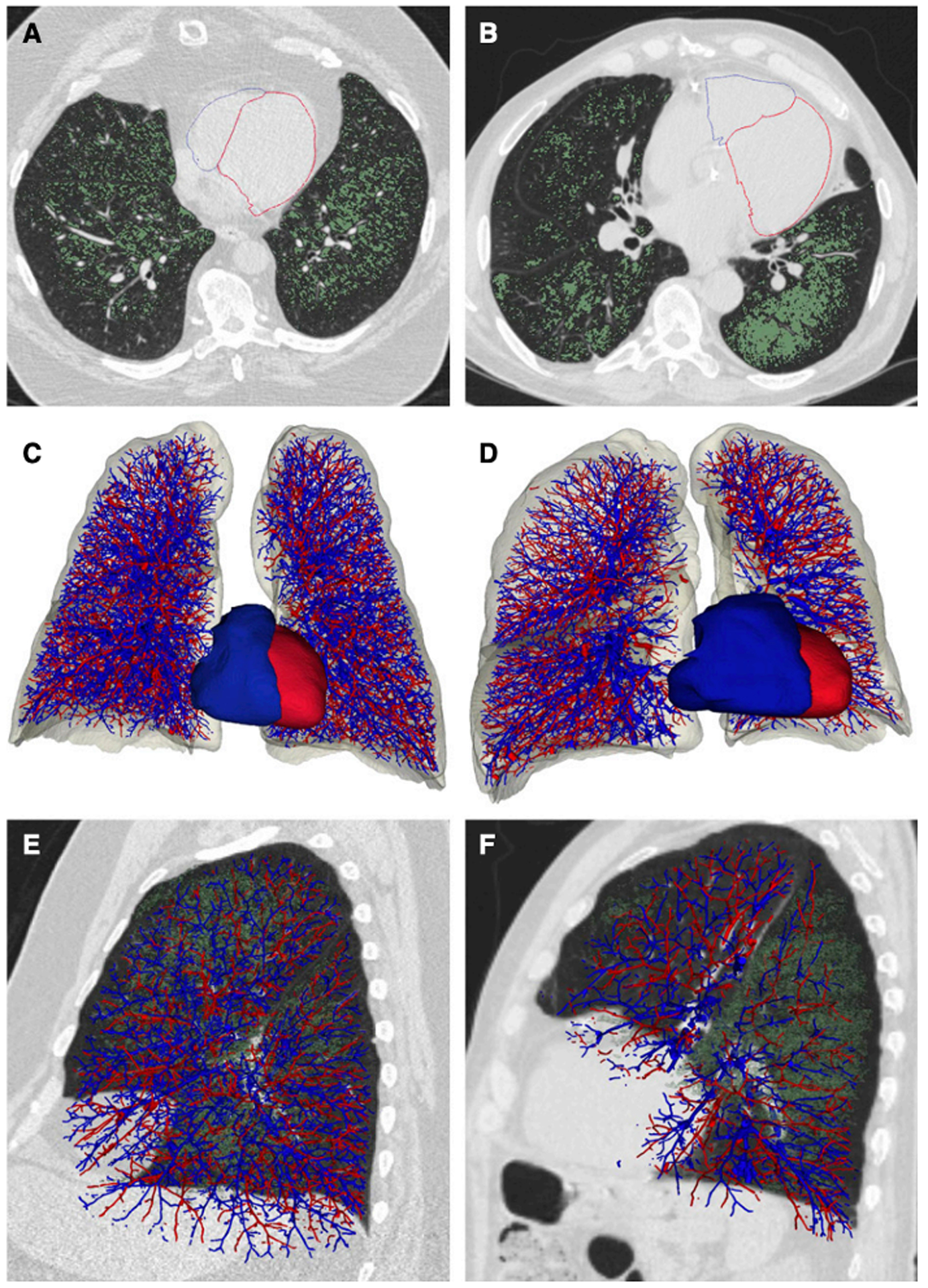
Pulmonary vasculature and right (blue) and left (red) ventricular reconstructions from computed tomography images for two subjects with approximately 20% emphysema on computed tomography scan. (A, C, and E) Subject 1 with 19% emphysema and relative preservation of the distal arterial vascular volume (arterial volume for vessels less than 5 mm2 in cross-section = 131 ml). (B, D, and F) Subject 2 with 18% emphysema and relative loss of the distal arterial vascular volume (arterial volume for vessels less than 5 mm2 in cross-section = 70.8 ml). (A and B) Axial images of the epicardial surface of the right ventricle (RV), which is outlined in blue and the epicardial surface of the left ventricle, which is outlined in red. Emphysema is depicted in green. (C and D) Frontal view of the arterial (blue) and venous vasculature and the surface model of the epicardial (myocardium and chamber) RV volume (blue) and epicardial (myocardium and chamber) left ventricular volume (red). The epicardial (myocardium and chamber) RV volume of Subject 1 is 58.9 ml and the epicardial (myocardium and chamber) RV volume for Subject 2 is 140 ml. (E and F) Sagittal views of the arterial (blue) and venous (red) vasculature of the left lung demonstrating the relative loss of distal arterial vascular volume. Emphysema is shown in green.
Reprinted with permission of the American Thoracic Society. Copyright © 2020 American Thoracic Society. American Journal of Respiratory and Critical Care Medicine 2019; 200(4): 454–461.
Skeletal Muscles
During the natural course of COPD, 15–40% of patients develop muscle wasting.41 A good measure that reflects skeletal muscle mass is the fat-free mass (FFM), and a low FFM is an independent risk factor for mortality in COPD.42 Traditional techniques used to measure FFM include bioelectrical impedance, skin-fold anthropometry, and dual X-ray absorptiometry. These three methods are not typically used in COPD clinics, whereas chest CT scanning is increasingly more common, particularly for lung cancer screening in heavy smokers. In the last few years, investigators have used chest CT imaging to evaluate muscle groups such as the pectoralis and paravertebral muscles.43–45 CT measurements of the pectoralis muscle area (PMA) are a reproducible measure of muscle mass and are easily obtained on a single axial slice.43,44 PMA correlates with bioelectrical impedance measures of muscle mass in healthy individuals and handgrip measures in subjects with COPD.46,47 Investigators have been able to derive FFM from CT measures of PMA, using two large cohorts of COPD subjects.48 The derived measure of FFM (i.e., FFMPMA) was then used to test the association between muscle mass and all-cause mortality. The investigators found that smokers with COPD and low FFMPMA had an increased risk of death.48 In smokers, muscle changes developed before they met the criteria for COPD. For instance, smokers without COPD have a lower oxidative fiber area and a higher glycolytic capacity than non-smoking controls. However, the prognostic value of muscle mass was unknown until recently.49 Investigators found that smokers without COPD in the lowest quartile of PMA (vs. the highest quartile) had an 85% higher risk of mortality.50 These findings highlight the ability of CT to provide an alternate assessment of skeletal muscle.
Bone Mineral Density
Osteoporosis is frequent in patients with COPD, and it is associated with an increased risk of bone fracture, which has prognostic implications. For instance, patients with COPD have a 60–70% increased risk of death following hip fracture compared to those without COPD.51 The potential causes of osteoporosis in this population include smoking, emphysema, lack of activity, steroid use, and chronic inflammation.52 The standard measure to diagnose osteoporosis is bone mineral density (BMD) using dual X-ray absorptiometry.52 Since CT imaging is based on determining tissue density, investigators have also used chest CT scanning to evaluate vertebral density. The method uses calcium calibration rods placed in the CT scanner pad under the subject and its density data stored within the chest CT image. A dedicated software determines the volumetric bone density —in mg/cc— (vBMD]) for each selected vertebra where a region of interest has been defined.53 Using this method, a study in over 3,200 smokers demonstrated that men with or without COPD (vs. their women counterparts) have a higher risk of low vBMD and vertebral fractures.53 The authors also showed that a higher burden of emphysema and airway disease on CT were each associated with low vBMD. In a short-term longitudinal study, it was found that a high burden of emphysema and greater levels of bone metabolism biomarkers (i.e., type I collagen C-telopeptide) also were associated with hip BMD decline at 2-year follow-up.54 Collectively, data on chest CT scanning applied to extra-pulmonary manifestations of COPD offers a unique opportunity to quantify structures concurrently, identify comorbidities (e.g., osteoporosis), and assess prognosis in both smokers at risk for and those already diagnosed with COPD.
COPD Progression Imaging
FEV1 change is the traditional measure of COPD progression. Large studies have demonstrated marked variability in FEV1 changes over time.55,56 The number of exacerbations, biomarker levels, such as Club cell protein 15, and CT measures of emphysema are factors associated with disease progression.55,57 In the last few years, advances in imaging have allowed for further insight into the pathological features and mechanical factors that contribute to disease progression. In a study in smokers with and without COPD, investigators used PRMfSAD and PRMEMPH to assess the relative importance of small airway disease or emphysema in predicting the decline in lung function.58 The investigators showed that PRMfSAD occurs in a significant number of subjects who do not meet the criteria for airflow limitation. While PRMfSAD but not PRMEMPH was significantly associated with FEV1 decline in subjects without COPD, both PRMfSAD and PRMEMPH were associated with FEV1 decline in GOLD 1–4 COPD subjects;58 however, the proportional contribution of the two CT metrics to the FEV1 decline relative to each other was higher for PRMfSAD than for PRMEMPH.58 This imaging technique has demonstrated that both small airway disease and emphysema are associated with disease progression. However, small airway disease seems more relevant in mild to moderate COPD, which has the highest rate of FEV1 decline. In another study, different disease processes were determined using factor analysis of 26 spirometric and CT variables. The loading of variables on the axes defined distinct disease processes. Two of these processes —airway-predominant axis and emphysema-predominant axis—resulted associated with increased mortality. The death risk varied based on the deciles of airway-predominant axis and emphysema-predominant axis in that the 9th and 10th deciles had the highest risk. These results allowed the investigators to create 6 groups of subjects as folllows:59 1) High-risk airway-predominant disease only; 2) Moderate-risk airway-predominant disease only; 3) High-risk emphysema-predominant disease only; 4) Combined high-risk airway- and emphysema-predominant disease; 5) Combined moderate-risk airway- and emphysema-predominant disease; and 6) No high-risk pulmonary group. The association between these groups and the GOLD stage progression was examined. The investigators found that the moderate-risk airway predominant disease was associated with the conversion of GOLD 0 to GOLD 2–4, while the high-risk emphysema-predominant disease was associated with the conversion of GOLD 0 to GOLD 1, and GOLD 1 to GOLD 4.59
Biomechanical stress is a conceivable mechanism of lung injury. Based on a proposed lung network model,60 initial damage to the alveolar walls (fibers) in emphysema concentrates stress on the remaining adjacent fibers. If local forces exceed fibers yield stress, then fibers can break, leading to the progression of the disease through direct mechanical injury. Imaging studies have now made clear how mechanically stress contributes to COPD progression. Investigators have assessed the mechanical stress of lung parenchyma using matched inspiratory-expiratory CT images. In matched CTs, the investigators measured regional changes of lung volume from total lung capacity (inspiratory CT) to functional residual capacity (expiratory CT), creating a deformation map representing lung areas of expansion and contraction. This metric is a Jacobian determinant, where values higher and lower than 1 indicate local expansion and contraction, respectively; a value of 1 indicates neither local expansion nor contraction (Figure 1). The investigators also assessed the Jacobian determinant on penumbras of a millimeter each around the emphysematous areas. In patients with COPD, they found that the Jacobian determinant was associated with FEV1/FVC and FEV1% predicted.61 They also demonstrated that the parenchyma within 2 mm of emphysematous areas was mechanically affected lung (MAL2) and called it “lung at risk.” They found that, in subjects with mild-to-moderate disease, the Jacobian determinant of MAL2 at or above vs. below the median value was associated with faster FEV1 decline (56.4 ml/yr vs. 43.2 ml/yr), supporting the long-recognized relevance of biomechanical stress in COPD progression.62
The prevailing view of the natural history of COPD proposed by Fletcher and Peto,63 that the rapid decline in FEV1 is caused by tobacco smoking, has been challenged.64 Only approximately half of the COPD subjects had a rapid FEV1 decline. In contrast, the other half never reached normal peak lung function in early adulthood and developed COPD in adulthood with an average rate of FEV1 decline.65 More recently, investigators have added CT phenotypic characteristics to the trajectories that can lead to COPD.66 First, lung function trajectory models were derived from a cohort of healthy men aged 21–80 years followed-up for a median of 29 years. These trajectories were then applied to a cohort of smokers, and their COPD phenotypes were compared. The subjects were scanned at two points in time, five years apart. The longitudinal analysis was performed by age (in years) groups, 45–55, 55–65, 65–75, and 75–85. The results demonstrated four distinct trajectories of lung function. 17% of subjects fell in the lowest trajectory (Trajectory 1), and they had the highest CT burden of emphysema and small airway disease across all age groups, whereas 36% fell in a low trajectory with evidence of increasing small airway disease and emphysema (Trajectory 2) with increasing age. The rest of the subjects fell in the remaining two trajectories (Trajectories 3 and 4) and tended to have preserved lung function and low burden of emphysema on CT (Figure 4). These findings and those of prior studies support the notions that lung development abnormalities may contribute to the development of COPD.64
Figure 4:
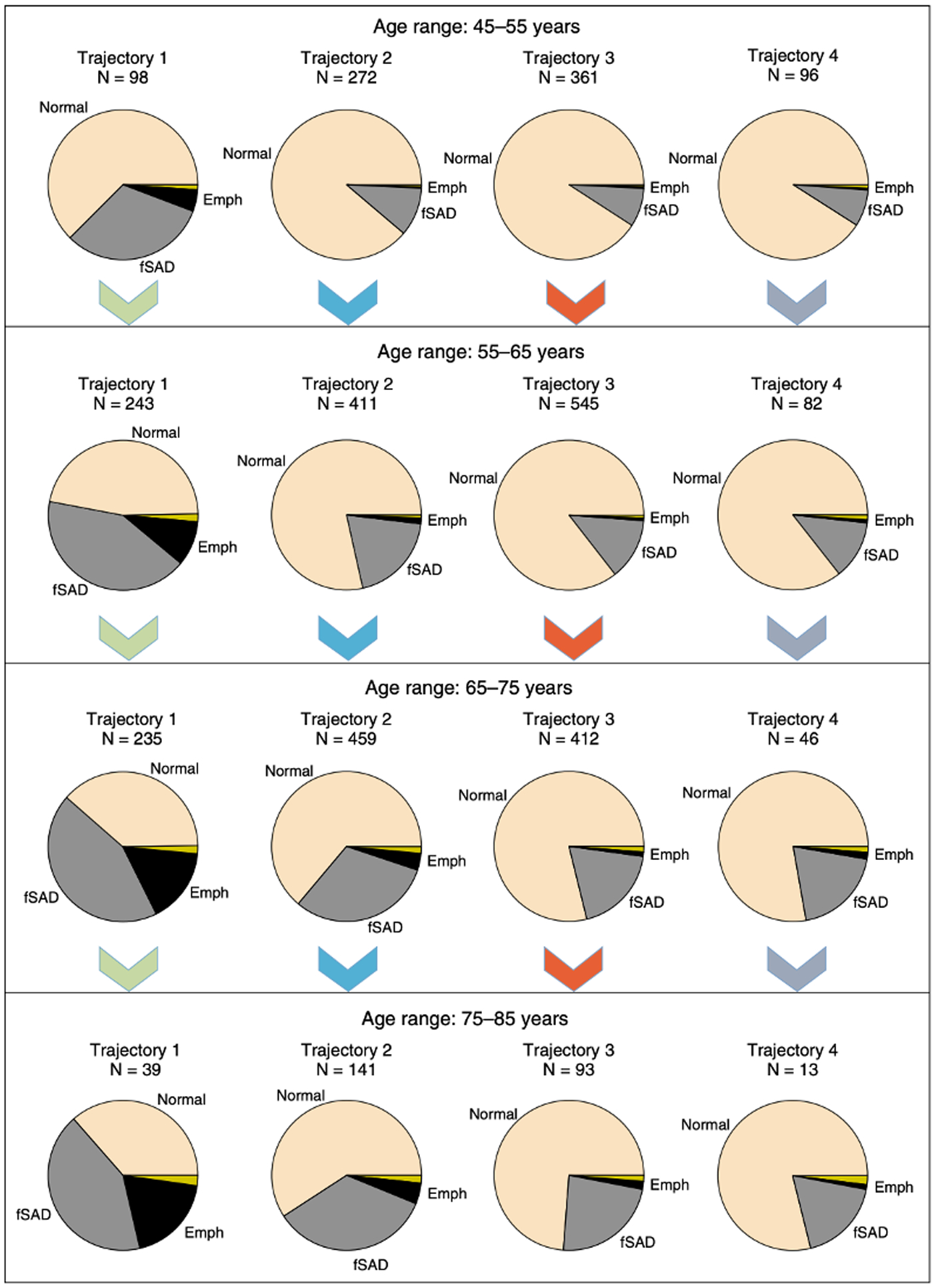
Age-stratified average relative composition of emphysema (Emph), functional small-airway disease (fSAD), normal tissue, and “other” (dark yellow portions) for each trajectory, as assessed by parametric response mapping analysis. Rows correspond to age strata, and columns correspond to trajectories; color-coded arrows clarify the direction of increasing age for each trajectory.
Reprinted with permission of the American Thoracic Society. Copyright © 2020 American Thoracic Society. American Journal of Respiratory and Critical Care Medicine 2018; 198(8):1033–1042.
In the last few years, imaging has also provided insight into the progression of COPD subtypes (i.e., emphysema and small airways disease predominant). Prior studies have suggested that small airways disease is a precursor of emphysema.1 The investigators have provided further evidence on this notion by demonstrating that baseline PRMfSAD is independently associated with emphysema at 5-yr follow-up.67 Other investigators have shown that the lobar distribution of emphysema is also related to different patterns of progression. In a cluster analysis study, investigators showed that the cluster most characterized by upper-lobe predominant emphysema at baseline experienced a faster overall progression of emphysema five years later.68 In the future, distinct patterns of COPD subtype progression may prove useful in the identification of patients for precision therapy; however, the impact of the radiographic changes on patient-specific outcomes still requires further investigation.
A few studies have used imaging as an end-point to evaluate therapy, particularly in alpha 1 anti-trypsin deficiency COPD to test anti-trypsin therapy replacement. In two consecutive clinical trials of augmentation therapy with an Alpha-1 Proteinase Inhibitor, the primary end-points were CT lung densities measured as TLC, functional residual capacity (FRC), and TLC+FRC combined.69,70 In the first trial, the investigators showed that the replacement therapy significantly slowed the progression of emphysema compared with placebo (mean annual rate of lung density loss of −1.45 g/L/year vs. −2.19 g/L/year).69 In the second trial, mean reduction in the yearly lung density between baseline and year 2, was significantly smaller in the early-start group (who received active therapy throughout), compared with the delayed-start group (who received placebo in the first trial; −1.51 g/L/year vs. −2.26 g/L/year).70 Those two trials also showed the therapeutic effect was more prominent in the basal region of the lung, highlighting that CT densitometry is a technically feasible outcome measure to assess the efficacy of emphysema-modifying therapies.71
Future Directions
While the imaging advances discussed here have enabled a richer understanding of COPD as a disease, there are still areas that require more progress. Chest imaging could prove to play a vital role in the clinical assessment of COPD, but further validation is required. Given the detailed information that CT imaging provides to identify COPD subtypes (e.g., emphysema); its role in the selection of patients for therapies such as lung volume reduction surgery or valve placement; and the improved quantification of disease progression; there is a compelling argument to be made for considering CT imaging as a more routine tool in the management of COPD. Although the findings of a few clinical trials using imaging as an endpoint are encouraging, more longitudinal imaging data is needed to provide better insight into disease subtype progression. Following this, there is great potential for the development of new therapies based on specific-disease subtypes.
A primary concern in the more routine use of CT imaging is radiation exposure. Although the current radiation exposure for chest CT is around 7.0 mSv, the further development of technologies to reduce this exposure level is desirable. Ongoing efforts to mitigate radiation exposure include automated exposure control, beam-shaping filters, and iterative reconstruction algorithms.
Conclusions
CT imaging is a robust in vivo tool that provides detailed information about changes in lung structures in subjects at risk for developing COPD, as well as about the subtype progression in those with COPD. Current advances in CT imaging are also proving useful for the examination of associated extra-pulmonary manifestations of the disease. Translations of these imaging findings into the development of new therapies for COPD is desirable.
Funding/Support:
Dr. Diaz receives funding support from the National Heart, Lung, and Blood Institute of the NIH under award number R01-HL133137 and the Brigham and Women’s Hospital Minority Faculty Career Development Award.
Footnotes
Conflict of Interest Disclosures:
Drs. Dolliver and Diaz have no conflicts of interest to disclose.
References
- 1.McDonough JE, Yuan R, Suzuki M, et al. Small-airway obstruction and emphysema in chronic obstructive pulmonary disease. N Engl J Med. 2011;365(17):1567–1575. [DOI] [PMC free article] [PubMed] [Google Scholar]
- 2.Diaz AA, Valim C, Yamashiro T, et al. Airway count and emphysema assessed by chest CT imaging predicts clinical outcome in smokers. Chest. 2010;138(4):880–887. [DOI] [PMC free article] [PubMed] [Google Scholar]
- 3.Kirby M, Tanabe N, Tan WC, et al. Total Airway Count on Computed Tomography and the Risk of Chronic Obstructive Pulmonary Disease Progression. Findings from a Population-based Study. Am J Respir Crit Care Med. 2018;197(1):56–65. [DOI] [PubMed] [Google Scholar]
- 4.Kirby M, Tanabe N, Vasilescu DM, et al. Computed Tomography Total Airway Count Is Associated with the Number of Micro-Computed Tomography Terminal Bronchioles. Am J Respir Crit Care Med. 2020;201(5):613–615. [DOI] [PubMed] [Google Scholar]
- 5.Regan EA, Hokanson JE, Murphy JR, et al. Genetic epidemiology of COPD (COPDGene) study design. COPD. 2010;7(1):32–43. [DOI] [PMC free article] [PubMed] [Google Scholar]
- 6.Vestbo J, Anderson W, Coxson HO, et al. Evaluation of COPD Longitudinally to Identify Predictive Surrogate End-points (ECLIPSE). Eur Respir J. 2008;31(4):869–873. [DOI] [PubMed] [Google Scholar]
- 7.Couper D, LaVange LM, Han M, et al. Design of the Subpopulations and Intermediate Outcomes in COPD Study (SPIROMICS). Thorax. 2014;69(5):491–494. [DOI] [PMC free article] [PubMed] [Google Scholar]
- 8.Bourbeau J, Tan WC, Benedetti A, et al. Canadian Cohort Obstructive Lung Disease (CanCOLD): Fulfilling the need for longitudinal observational studies in COPD. COPD. 2014;11(2):125–132. [DOI] [PubMed] [Google Scholar]
- 9.Bodduluri S, Reinhardt JM, Hoffman EA, Newell JD Jr., Bhatt SP. Recent Advances in Computed Tomography Imaging in Chronic Obstructive Pulmonary Disease. Annals of the American Thoracic Society. 2018;15(3):281–289. [DOI] [PMC free article] [PubMed] [Google Scholar]
- 10.Thiboutot J, Yuan W, Park HC, et al. Current Advances in COPD Imaging. Academic radiology. 2019;26(3):335–343. [DOI] [PMC free article] [PubMed] [Google Scholar]
- 11.Singh D, Agusti A, Anzueto A, et al. Global Strategy for the Diagnosis, Management, and Prevention of Chronic Obstructive Lung Disease: the GOLD science committee report 2019. Eur Respir J. 2019;53(5). [DOI] [PubMed] [Google Scholar]
- 12.Regan EA, Lynch DA, Curran-Everett D, et al. Clinical and Radiologic Disease in Smokers With Normal Spirometry. JAMA Intern Med. 2015;175(9):1539–1549. [DOI] [PMC free article] [PubMed] [Google Scholar]
- 13.Colak Y, Nordestgaard BG, Vestbo J, Lange P, Afzal S. Prognostic significance of chronic respiratory symptoms in individuals with normal spirometry. Eur Respir J. 2019;54(3). [DOI] [PubMed] [Google Scholar]
- 14.Woodruff PG, Barr RG, Bleecker E, et al. Clinical Significance of Symptoms in Smokers with Preserved Pulmonary Function. N Engl J Med. 2016;374(19):1811–1821. [DOI] [PMC free article] [PubMed] [Google Scholar]
- 15.Bowler RP, Kim V, Regan E, et al. Prediction of acute respiratory disease in current and former smokers with and without COPD. Chest. 2014;146(4):941–950. [DOI] [PMC free article] [PubMed] [Google Scholar]
- 16.Washko GR, Kinney GL, Ross JC, et al. Lung Mass in Smokers. Academic radiology. 2017;24(4):386–392. [DOI] [PMC free article] [PubMed] [Google Scholar]
- 17.Harmouche R, Ash SY, Putman RK, et al. Objectively Measured Chronic Lung Injury on Chest CT. Chest. 2019;156(6):1149–1159. [DOI] [PMC free article] [PubMed] [Google Scholar]
- 18.Oelsner EC, Smith BM, Hoffman EA, et al. Associations between emphysema-like lung on CT and incident airflow limitation: a general population-based cohort study. Thorax. 2018;73(5):486–488. [DOI] [PMC free article] [PubMed] [Google Scholar]
- 19.Washko GR, Diaz AA, Kim V, et al. Computed tomographic measures of airway morphology in smokers and never-smoking normals. J Appl Physiol (1985). 2014;116(6):668–673. [DOI] [PMC free article] [PubMed] [Google Scholar]
- 20.Diaz AA, Rahaghi FN, Ross JC, et al. Understanding the contribution of native tracheobronchial structure to lung function: CT assessment of airway morphology in never smokers. Respir Res. 2015;16:23. [DOI] [PMC free article] [PubMed] [Google Scholar]
- 21.Smith BM, Hoffman EA, Rabinowitz D, et al. Comparison of spatially matched airways reveals thinner airway walls in COPD. The Multi-Ethnic Study of Atherosclerosis (MESA) COPD Study and the Subpopulations and Intermediate Outcomes in COPD Study (SPIROMICS). Thorax. 2014;69(11):987–996. [DOI] [PMC free article] [PubMed] [Google Scholar]
- 22.Smith BM, Kirby M, Hoffman EA, et al. Association of Dysanapsis With Chronic Obstructive Pulmonary Disease Among Older Adults. Jama. 2020;323(22):2268–2280. [DOI] [PMC free article] [PubMed] [Google Scholar]
- 23.Galban CJ, Han MK, Boes JL, et al. Computed tomography-based biomarker provides unique signature for diagnosis of COPD phenotypes and disease progression. Nat Med. 2012;18(11):1711–1715. [DOI] [PMC free article] [PubMed] [Google Scholar]
- 24.Vasilescu DM, Martinez FJ, Marchetti N, et al. Noninvasive Imaging Biomarker Identifies Small Airway Damage in Severe Chronic Obstructive Pulmonary Disease. Am J Respir Crit Care Med. 2019;200(5):575–581. [DOI] [PMC free article] [PubMed] [Google Scholar]
- 25.Boudewijn IM, Postma DS, Telenga ED, et al. Effects of ageing and smoking on pulmonary computed tomography scans using parametric response mapping. Eur Respir J. 2015;46(4):1193–1196. [DOI] [PMC free article] [PubMed] [Google Scholar]
- 26.Smith BM, Traboulsi H, Austin JHM, et al. Human airway branch variation and chronic obstructive pulmonary disease. Proc Natl Acad Sci U S A. 2018;115(5):E974–E981. [DOI] [PMC free article] [PubMed] [Google Scholar]
- 27.Koo HK, Vasilescu DM, Booth S, et al. Small airways disease in mild and moderate chronic obstructive pulmonary disease: a cross-sectional study. Lancet Respir Med. 2018;6(8):591–602. [DOI] [PubMed] [Google Scholar]
- 28.Diaz AA. The Case of Missing Airways in Chronic Obstructive Pulmonary Disease. Am J Respir Crit Care Med. 2018;197(1):4–6. [DOI] [PMC free article] [PubMed] [Google Scholar]
- 29.Nathan SD, Barbera JA, Gaine SP, et al. Pulmonary hypertension in chronic lung disease and hypoxia. Eur Respir J. 2019;53(1). [DOI] [PMC free article] [PubMed] [Google Scholar]
- 30.Iyer AS, Wells JM, Vishin S, Bhatt SP, Wille KM, Dransfield MT. CT scan-measured pulmonary artery to aorta ratio and echocardiography for detecting pulmonary hypertension in severe COPD. Chest. 2014;145(4):824–832. [DOI] [PMC free article] [PubMed] [Google Scholar]
- 31.Wells JM, Iyer AS, Rahaghi FN, et al. Pulmonary artery enlargement is associated with right ventricular dysfunction and loss of blood volume in small pulmonary vessels in chronic obstructive pulmonary disease. Circ Cardiovasc Imaging. 2015;8(4). [DOI] [PMC free article] [PubMed] [Google Scholar]
- 32.Cuttica MJ, Bhatt SP, Rosenberg SR, et al. Pulmonary artery to aorta ratio is associated with cardiac structure and functional changes in mild-to-moderate COPD. International journal of chronic obstructive pulmonary disease. 2017;12:1439–1446. [DOI] [PMC free article] [PubMed] [Google Scholar]
- 33.Wells JM, Washko GR, Han MK, et al. Pulmonary arterial enlargement and acute exacerbations of COPD. N Engl J Med. 2012;367(10):913–921. [DOI] [PMC free article] [PubMed] [Google Scholar]
- 34.LaFon DC, Bhatt SP, Labaki WW, et al. Pulmonary artery enlargement and mortality risk in moderate to severe COPD: results from COPDGene. Eur Respir J. 2020;55(2). [DOI] [PMC free article] [PubMed] [Google Scholar]
- 35.Shin S, King CS, Brown AW, et al. Pulmonary artery size as a predictor of pulmonary hypertension and outcomes in patients with chronic obstructive pulmonary disease. Respiratory medicine. 2014;108(11):1626–1632. [DOI] [PubMed] [Google Scholar]
- 36.Terzikhan N, Bos D, Lahousse L, et al. Pulmonary artery to aorta ratio and risk of all-cause mortality in the general population: the Rotterdam Study. Eur Respir J. 2017;49(6). [DOI] [PubMed] [Google Scholar]
- 37.Grau M, Barr RG, Lima JA, et al. Percent emphysema and right ventricular structure and function: the Multi-Ethnic Study of Atherosclerosis-Lung and Multi-Ethnic Study of Atherosclerosis-Right Ventricle Studies. Chest. 2013;144(1):136–144. [DOI] [PMC free article] [PubMed] [Google Scholar]
- 38.Weitzenblum E Chronic cor pulmonale. Heart. 2003;89(2):225–230. [DOI] [PMC free article] [PubMed] [Google Scholar]
- 39.Washko GR, Nardelli P, Ash SY, et al. Arterial Vascular Pruning, Right Ventricular Size, and Clinical Outcomes in Chronic Obstructive Pulmonary Disease. A Longitudinal Observational Study. Am J Respir Crit Care Med. 2019;200(4):454–461. [DOI] [PMC free article] [PubMed] [Google Scholar]
- 40.Rahaghi FN, Vegas-Sanchez-Ferrero G, Minhas JK, et al. Ventricular Geometry From Non-contrast Non-ECG-gated CT Scans: An Imaging Marker of Cardiopulmonary Disease in Smokers. Academic radiology. 2017;24(5):594–602. [DOI] [PMC free article] [PubMed] [Google Scholar]
- 41.Sanders KJ, Kneppers AE, van de Bool C, Langen RC, Schols AM. Cachexia in chronic obstructive pulmonary disease: new insights and therapeutic perspective. J Cachexia Sarcopenia Muscle. 2016;7(1):5–22. [DOI] [PMC free article] [PubMed] [Google Scholar]
- 42.Schols AM, Broekhuizen R, Weling-Scheepers CA, Wouters EF. Body composition and mortality in chronic obstructive pulmonary disease. Am J Clin Nutr. 2005;82(1):53–59. [DOI] [PubMed] [Google Scholar]
- 43.McDonald ML, Diaz AA, Ross JC, et al. Quantitative computed tomography measures of pectoralis muscle area and disease severity in chronic obstructive pulmonary disease. A cross-sectional study. Annals of the American Thoracic Society. 2014;11(3):326–334. [DOI] [PMC free article] [PubMed] [Google Scholar]
- 44.Diaz AA, Zhou L, Young TP, et al. Chest CT measures of muscle and adipose tissue in COPD: gender-based differences in content and in relationships with blood biomarkers. Academic radiology. 2014;21(10):1255–1261. [DOI] [PMC free article] [PubMed] [Google Scholar]
- 45.Tanimura K, Sato S, Fuseya Y, et al. Quantitative Assessment of Erector Spinae Muscles in Patients with Chronic Obstructive Pulmonary Disease. Novel Chest Computed Tomography-derived Index for Prognosis. Annals of the American Thoracic Society. 2016;13(3):334–341. [DOI] [PubMed] [Google Scholar]
- 46.Kim YS, Kim EY, Kang SM, Ahn HK, Kim HS. Single cross-sectional area of pectoralis muscle by computed tomography - correlation with bioelectrical impedance based skeletal muscle mass in healthy subjects. Clin Physiol Funct Imaging. 2017;37(5):507–511. [DOI] [PubMed] [Google Scholar]
- 47.Martinez CH, Diaz AA, Meldrum CA, et al. Handgrip Strength in Chronic Obstructive Pulmonary Disease. Associations with Acute Exacerbations and Body Composition. Annals of the American Thoracic Society. 2017;14(11):1638–1645. [DOI] [PMC free article] [PubMed] [Google Scholar]
- 48.McDonald MN, Diaz AA, Rutten E, et al. Chest computed tomography-derived low fat-free mass index and mortality in COPD. Eur Respir J. 2017;50(6). [DOI] [PMC free article] [PubMed] [Google Scholar]
- 49.Montes de Oca M, Loeb E, Torres SH, De Sanctis J, Hernandez N, Talamo C. Peripheral muscle alterations in non-COPD smokers. Chest. 2008;133(1):13–18. [DOI] [PubMed] [Google Scholar]
- 50.Diaz AA, Martinez CH, Harmouche R, et al. Pectoralis muscle area and mortality in smokers without airflow obstruction. Respir Res. 2018;19(1):62. [DOI] [PMC free article] [PubMed] [Google Scholar]
- 51.de Luise C, Brimacombe M, Pedersen L, Sorensen HT. Chronic obstructive pulmonary disease and mortality following hip fracture: a population-based cohort study. Eur J Epidemiol. 2008;23(2):115–122. [DOI] [PubMed] [Google Scholar]
- 52.Bon J, Fuhrman CR, Weissfeld JL, et al. Radiographic emphysema predicts low bone mineral density in a tobacco-exposed cohort. Am J Respir Crit Care Med. 2011;183(7):885–890. [DOI] [PMC free article] [PubMed] [Google Scholar]
- 53.Jaramillo JD, Wilson C, Stinson DS, et al. Reduced Bone Density and Vertebral Fractures in Smokers. Men and COPD Patients at Increased Risk. Annals of the American Thoracic Society. 2015;12(5):648–656. [DOI] [PMC free article] [PubMed] [Google Scholar]
- 54.Bon J, Zhang Y, Leader JK, et al. Radiographic Emphysema, Circulating Bone Biomarkers, and Progressive Bone Mineral Density Loss in Smokers. Annals of the American Thoracic Society. 2018;15(5):615–621. [DOI] [PMC free article] [PubMed] [Google Scholar]
- 55.Vestbo J, Edwards LD, Scanlon PD, et al. Changes in forced expiratory volume in 1 second over time in COPD. N Engl J Med. 2011;365(13):1184–1192. [DOI] [PubMed] [Google Scholar]
- 56.Diaz AA, Strand M, Coxson HO, et al. Disease Severity Dependence of the Longitudinal Association Between CT Lung Density and Lung Function in Smokers. Chest. 2018;153(3):638–645. [DOI] [PMC free article] [PubMed] [Google Scholar]
- 57.Coxson HO, Dirksen A, Edwards LD, et al. The presence and progression of emphysema in COPD as determined by CT scanning and biomarker expression: a prospective analysis from the ECLIPSE study. Lancet Respir Med. 2013;1(2):129–136. [DOI] [PubMed] [Google Scholar]
- 58.Bhatt SP, Soler X, Wang X, et al. Association between Functional Small Airway Disease and FEV1 Decline in Chronic Obstructive Pulmonary Disease. Am J Respir Crit Care Med. 2016;194(2):178–184. [DOI] [PMC free article] [PubMed] [Google Scholar]
- 59.Young KA, Strand M, Ragland MF, et al. Pulmonary Subtypes Exhibit Differential Global Initiative for Chronic Obstructive Lung Disease Spirometry Stage Progression: The COPDGene(R) Study. Chronic Obstr Pulm Dis. 2019;6(5):414–429. [DOI] [PMC free article] [PubMed] [Google Scholar]
- 60.Ingenito EP, Tsai LW, Majumdar A, Suki B. On the role of surface tension in the pathophysiology of emphysema. Am J Respir Crit Care Med. 2005;171(4):300–304. [DOI] [PubMed] [Google Scholar]
- 61.Bhatt SP, Bodduluri S, Newell JD, et al. CT-derived Biomechanical Metrics Improve Agreement Between Spirometry and Emphysema. Academic radiology. 2016;23(10):1255–1263. [DOI] [PMC free article] [PubMed] [Google Scholar]
- 62.Bhatt SP, Bodduluri S, Hoffman EA, et al. Computed Tomography Measure of Lung at Risk and Lung Function Decline in Chronic Obstructive Pulmonary Disease. Am J Respir Crit Care Med. 2017;196(5):569–576. [DOI] [PMC free article] [PubMed] [Google Scholar]
- 63.Fletcher C, Peto R. The natural history of chronic airflow obstruction. Br Med J. 1977;1(6077):1645–1648. [DOI] [PMC free article] [PubMed] [Google Scholar]
- 64.Agusti A, Faner R. The Changing Landscape of Chronic Obstructive Pulmonary Disease. Am J Respir Crit Care Med. 2018;198(8):978–981. [DOI] [PubMed] [Google Scholar]
- 65.Lange P, Celli B, Agusti A, et al. Lung-Function Trajectories Leading to Chronic Obstructive Pulmonary Disease. N Engl J Med. 2015;373(2):111–122. [DOI] [PubMed] [Google Scholar]
- 66.Ross JC, Castaldi PJ, Cho MH, et al. Longitudinal Modeling of Lung Function Trajectories in Smokers with and without Chronic Obstructive Pulmonary Disease. Am J Respir Crit Care Med. 2018;198(8):1033–1042. [DOI] [PMC free article] [PubMed] [Google Scholar]
- 67.Labaki WW, Gu T, Murray S, et al. Voxel-Wise Longitudinal Parametric Response Mapping Analysis of Chest Computed Tomography in Smokers. Academic radiology. 2019;26(2):217–223. [DOI] [PMC free article] [PubMed] [Google Scholar]
- 68.Boueiz A, Chang Y, Cho MH, et al. Lobar Emphysema Distribution Is Associated With 5-Year Radiological Disease Progression. Chest 2018;153(1):65–76. [DOI] [PMC free article] [PubMed] [Google Scholar]
- 69.Chapman KR, Burdon JG, Piitulainen E, et al. Intravenous augmentation treatment and lung density in severe alpha1 antitrypsin deficiency (RAPID): a randomised, double-blind, placebo-controlled trial. Lancet. 2015;386(9991):360–368. [DOI] [PubMed] [Google Scholar]
- 70.McElvaney NG, Burdon J, Holmes M, et al. Long-term efficacy and safety of alpha1 proteinase inhibitor treatment for emphysema caused by severe alpha1 antitrypsin deficiency: an open-label extension trial (RAPID-OLE). Lancet Respir Med. 2017;5(1):51–60. [DOI] [PubMed] [Google Scholar]
- 71.Campos MA, Diaz AA. The Role of Computed Tomography for the Evaluation of Lung Disease in Alpha-1 Antitrypsin Deficiency. Chest. 2018;153(5):1240–1248. [DOI] [PMC free article] [PubMed] [Google Scholar]