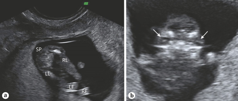
Fig. 1.
Ultrasound images at the 13th week of pregnancy. a Both lower limbs of the fetus. Note mesomelic shortening and malposition of both feet. b Coronal view of the fetal facies. The arrows point to both crystalline lens, showing pronounced hypertelorism. SP, spine; LL, left leg; RL, right leg; LF, left foot; RF, right foot.

