. 2020 May 29;2(3):e190076. doi: 10.1148/rycan.2020190076

Figure 3:

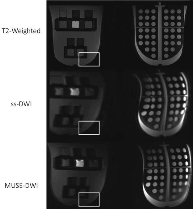
Axial images from phantom testing. T2-weighted image (top panel), single-shot diffusion-weighted image (ss-DWI) (middle panel), and high-spatial-resolution multiplexed sensitivity-encoding diffusion weighted image (MUSE-DWI) with protocol D (bottom panel) are shown. The region of interest is placed to show the presence of geometric distortion artifact on diffusion-weighted images, which is partially corrected with MUSE DWI.

Axial images from phantom testing. T2-weighted image (top panel), single-shot diffusion-weighted image (ss-DWI) (middle panel), and high-spatial-resolution multiplexed sensitivity-encoding diffusion weighted image (MUSE-DWI) with protocol D (bottom panel) are shown. The region of interest is placed to show the presence of geometric distortion artifact on diffusion-weighted images, which is partially corrected with MUSE DWI.