. 2020 May 29;2(3):e190076. doi: 10.1148/rycan.2020190076

Figure 5:

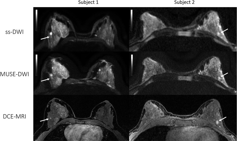
Axial images from two patients with benign lesions. Left: Patient 1, a 43-year-old woman with a 5-mm enhancing focus in the outer right breast in which stability was verified after 2-year follow-up (arrow). Right: Patient 2, a 47-year-old woman with an enhancing 7-mm nodule in the lower inner left quadrant (arrow). Biopsy results revealed fibroadenoma. Multiplexed sensitivity-encoding diffusion-weighted imaging (MUSE-DWI) yielded a sharper delineation of the breast parenchyma, and lesion contours appear to be more defined than in single-shot DWI (b value, 800 sec/mm2) (ss-DWI). These two examples were categorized as 1 (better overall image quality with MUSE DWI than with single-shot DWI) by both readers. DCE-MRI = dynamic contrast-enhanced MRI.

Axial images from two patients with benign lesions. Left: Patient 1, a 43-year-old woman with a 5-mm enhancing focus in the outer right breast in which stability was verified after 2-year follow-up (arrow). Right: Patient 2, a 47-year-old woman with an enhancing 7-mm nodule in the lower inner left quadrant (arrow). Biopsy results revealed fibroadenoma. Multiplexed sensitivity-encoding diffusion-weighted imaging (MUSE-DWI) yielded a sharper delineation of the breast parenchyma, and lesion contours appear to be more defined than in single-shot DWI (b value, 800 sec/mm2) (ss-DWI). These two examples were categorized as 1 (better overall image quality with MUSE DWI than with single-shot DWI) by both readers. DCE-MRI = dynamic contrast-enhanced MRI.