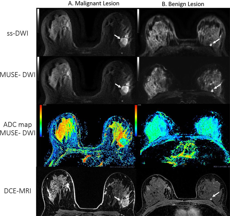
Figure 7:
Axial single-shot diffusion-weighted images (ss-DWI), images from multiplexed sensitivity-encoding DWI (MUSE-DWI), apparent diffusion coefficient (ADC) maps, and dynamic contrast-enhanced MRI (DCE-MRI) images from two patients with lesions (white arrows). A, Images in a 27-year-old woman with a 25-mm irregular heterogeneously enhancing mass in the left breast. A satellite nodule is evident anterior to the main lesion. At biopsy, the lesion was identified as invasive ductal carcinoma. Readers considered the overall image quality for this case to be worse (category 3) with MUSE DWI than with single-shot DWI. B, A 55-year-old woman with a history of right breast cancer after right lumpectomy. A 6-mm enhancing lesion is seen in the posterior third of the left breast (arrow), which was unchanged for the previous 2 years. Both readers scored a better overall image quality (category 1) with MUSE DWI for this case. ADC values are measured in × 10−6 mm2/sec for both images. ADC values obtained from the region of interest (ROI) in, A: mean, 793.7 × 10⁻⁶ mm2/sec; minimum, 13 × 10–6 mm2/sec; maximum, 1431 × 10–6 mm2/sec; region of interest area, 57.4 mm2. ADC values obtained from the ROI in, B: mean, 2200 × 10–6 mm2/sec; minimum, 1368 × 10−6 mm2/sec; maximum, 2928 × 10−6 mm2/sec; region of interest area, 41.2 mm2. Blue indicates lower ADC value, red indicates higher ADC value. Note that the minimum ADC threshold value in the color bar for lesion A is 0,whereas in the color bar for lesion B it is 1062 to enhance visibility of this benign lesion.

