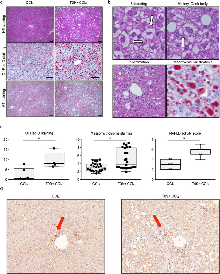
Figure 2.

Histopathological features and typical scores of non‐alcoholic steatohepatitis. (a) Hematoxylin and eosin (HE), Oil Red O, and Masson's trichrome (MT) staining of tissues from representative mice in the CCl4 and T09 + CCl4 groups. Scale bar = 100 μm. (b) Representative HE staining of liver tissue from the T09 + CCl4 group depicting the individual components of steatohepatitis—ballooning, Mallory–Denk bodies, inflammation, and macrovesicular steatosis. (c) Quantification of histological scores for steatosis, fibrosis, and non‐alcoholic fatty liver disease (NAFLD) activity. The results are expressed as the mean ± SD and were compared with the Mann–Whitney U‐test. (a) Oil Red O staining (n = 5). (b) MT staining (n = 5). (c) NAFLD activity score (n = 7). (d) Representative 4‐HNE immunostaining of liver tissue from the T09 + CCl4 and CCl4 groups. [Color figure can be viewed at wileyonlinelibrary.com] [Color figure can be viewed at wileyonlinelibrary.com]
