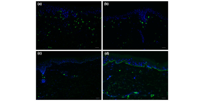
Figure 1.

Immunofluorescent staining of IgE in the skin of bullous pemphigoid (BP) patients. (a) Many IgE‐positive cells in the dermis (++), representative for five of 14 BP skin samples. (b) Multiple IgE‐positive cells in the dermis (+), representative for one of 14 BP skin samples. (c) Few IgE‐positive cells in the dermis (+/−), representative for six of 14 BP skin samples. (d) One BP skin sample showed linear IgE along the basement membrane zone, while 13 other skin samples did not. White bar is 50 μm.
