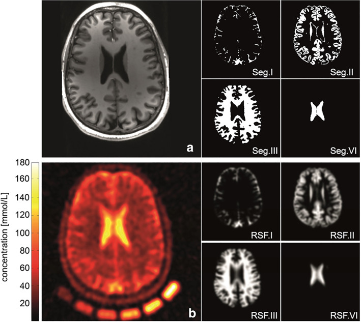
FIGURE 2.

Anatomical information (a) coregistered to quantitative sodium image (b) normalized by means of the external calibration cushion (nominal spatial resolution: (3 mm3), 3600 projections, TR = 200 msec, TE = 0.35 msec; a Hamming‐filter was used to reduce Gibbs ringing artifacts and to gain SNR (reduced shown FOV of 220 × 220 × 220 mm3). MPRAGE (a) and a CISS sequence were acquired to derive tissue masks. Segmentation was performed using the FSL toolkit (Jenkinson and Smith, 2001). The cerebrospinal fluid (CSF) volume was divided into two separate compartments to enable an intrinsic correction control of the performed PVC. Example slices of used 3D‐segmentation masks used in the correction algorithm: (Seg.I) of outer CSF, (Seg.VI) inner CSF volume, (Seg.II) gray matter, and (Seg.II) white matter. Corresponding region spread function (RSFs) for the single compartments (RSF.I–RSF.IV). The calculated broadening of the single compartments is in good agreement with the observed blurring in the measured sodium image. Reproduced from Ref. 39 with permission from Elsevier, Neuroimage.
